Using AI to identify high-risk patients in home health care

The population is aging. Over the past two decades, life expectancy in the United States has increased by more than five years to approximately 80 years in 2020 and is projected to further increase to over 85 years in 2060. The progressive rise in life expectancy leads to a growing share of older people in society, which increases the demand for health care services including home health care (HHC). HHC is defined by medical services that are provided in a patient’s home, usually by skilled nurses. An important aspect of HHC is the identification of patients who are at high risk for emergency care. A substantial fraction of HHC patients has to visit the emergency room or is admitted to the hospital during the HHC period. Strikingly, up to 30% of these events could be avoided by accurate risk prediction and preventative care. For example, patients identified as high-risk can be monitored more closely and treated with additional medications to prevent adverse health outcomes.

The research of Columbia postdoc Jiyoun Song is aimed at improving risk prediction in the setting of HHC. In recent work published in the Journal of Advanced Nursing, Dr. Song and colleagues performed cluster analysis, an unsupervised machine learning method to aggregate available patient data into groups. Such a clustering method is useful in identifying patterns of risk factors that interact with each other, rather than examining individual risk factors. The analysis was performed on structured data from electronic health records, that include patient characteristics such as socio-demographics and health conditions, as well as unstructured data from clinical notes written by nurses. The approach of Dr. Song and colleagues to use data derived from these clinical notes is quite innovative. They extracted risk factors from these notes through the artificial intelligence (AI)-based tool of natural language processing. Natural language processing can be used to extract meaning from text written by a human, which forms the foundation of AI chatbot systems such as the recently developed ChatGTP. The use of clinical notes as an additional source of data significantly improves risk prediction in the HHC setting, as previously shown by Dr. Song.

Through AI-assisted cluster analysis of both the structured and unstructured data, three clusters of risk factors were identified with distinct characteristics: (1) impaired physical comfort with pain, (2) high comorbidity burden (i.e., a person is suffering from more than one physical disease or condition at the same time), and (3) impaired cognitive/psychological and skin integrity. Classifying patients in these three categories could help tailor individualized preventive strategies. For example, a pain management strategy may work best for patients in cluster 1, while patients in cluster 3 may benefit mostly from psychological counseling and wound management. The study also found that these clusters are associated with the frequency and timing of emergency room visits. Patients that fall into cluster 3 have the highest need for emergency care, with 15.7% of patients being hospitalized or visiting the emergency department within the first 60 days of HHC (see Figure below).

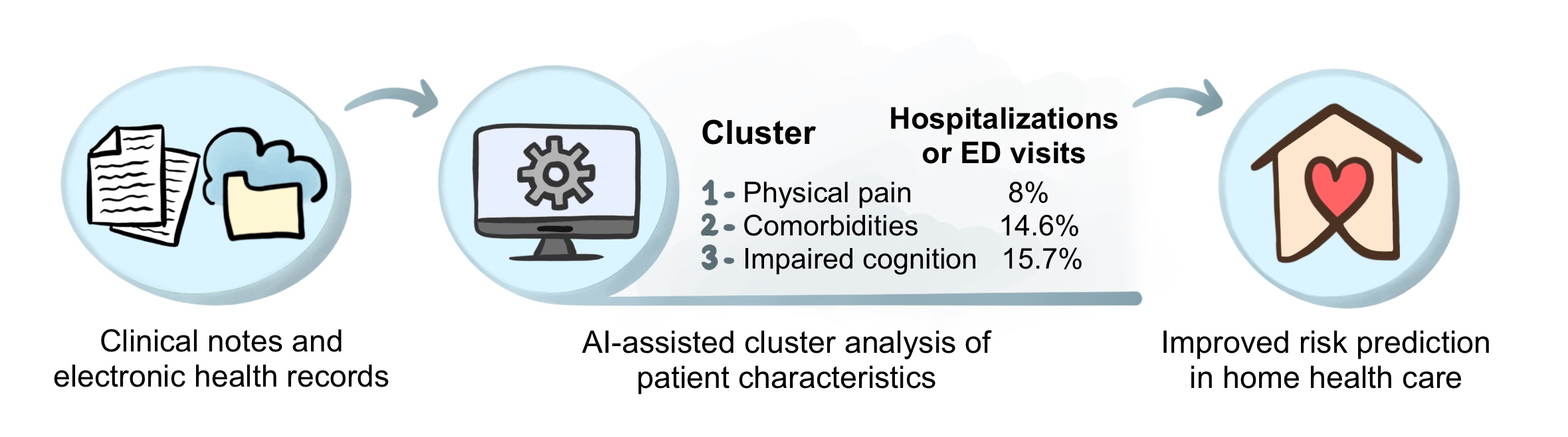
AI-assisted cluster analysis of both structured data (i.e., electronic health records) and unstructured data (i.e., clinical notes) identified three clusters of risk factors with distinct characteristics. The risk of hospitalizations or emergency department (ED) visits is different for each cluster. Such a cluster-based analysis is useful for identifying high-risk patients in home health care and implementing preventative strategies. © 2022, Maaike Schilperoort

The results from this study suggest that implementation of cluster-based risk prediction models into early warning systems could reduce the likelihood of HHC patients being admitted to the hospital. This illustrates the potential of AI-based methods for clinical risk prediction. The use of AI is getting increasingly popular and is now also implemented in various other aspects of healthcare, such as in-hospital decision making, predicting treatment benefit, and personalizing medicine. To what extent will AI be incorporated in our healthcare system? Only time will tell.

Reviewed by: Jiyoun Song, Pei-Yin Shih, Trang Nguyen, and Sam Rossano

A how-to guide for improving the potency of stem cells

You may remember Dolly, the sheep that became famous in the ‘90s as the first mammal to be cloned from an adult cell. Dolly was created through somatic cell nuclear transfer (SCNT), in which the nucleus from a somatic donor cell, i.e., a cell from the body other than a sperm or egg cell, is transferred into an enucleated egg cell. In this case, the donor cell was derived from a sheep’s mammary glands, a medical term for the breasts. The scientists named the cloned sheep Dolly since they could not think of a more impressive pair of mammary glands than Dolly Parton’s, or so the story goes. Aside from generating viable embryos in the laboratory, SCNT can be used to generate human stem cell lines for research and therapeutic purposes. However, this procedure is technically challenging and requires egg cells, which raises ethical concerns.

Artist’s impression of Dolly Parton, the famous American country singer, holding the cloned sheep named after her.
© 2022, Maaike Schilperoort

In 2007, a lab in Kyoto, Japan, found another way of generating human stem cells. The group infected human skin cells with a virus that carried a set of genes known to be important for embryonic stem cells. This resulted in so-called “induced pluripotent stem cells”, or iPSCs, that are functionally identical to embryonic stem cells. Although therapeutically promising, these iPSCs do not have the same potency as the cells generated through SCNT. SCNT generates cells that are totipotent at an early stage, meaning that they can form viable embryos as well as extraembryonic tissues such as the placenta and yolk sack. In contrast, iPSCs are pluripotent and are not able to give rise to extraembryonic tissues. They also have an inferior differentiation potential and lower proliferation rate as compared to totipotent cells.

Efforts have been made by scientists to make embryonic stem cells and iPSCs more totipotent by treating them with small molecule inhibitors, resulting in so-called expanded potential stem cells (EPSCs) that that can give rise to the embryo as well as placenta tissues and thus are more versatile as compared to their pluripotent counterparts. However, the developmental potential of EPSCs is still inferior to true totipotent cells or cells generated through SCNT. To gain insight into how the developmental potential of EPSCs can be improved, Columbia postdoc Vikas Malik and colleagues performed a deep analysis of pluripotent embryonic stem cells vs. the more totipotent EPSCs. They examined gene expression, DNA accessibility, and protein expression, and found some unique genes and proteins that are upregulated in EPSCs as compared to embryonic stem cells, such as Zscan4c, Rara, Zfp281, and UTF1. This pioneering work, published in Life Science Alliance, shows us which genes and proteins to target to generate authentic totipotent stem cells in a petri dish.

The work of Dr. Malik and colleagues has improved our understanding of how to generate totipotent cells outside of the human body without having to deal with the technical and ethical challenges of SCNT. These cells can further improve stem cell therapy through a greater ability to regenerate and repair tissues affected by damage or disease. In addition, totipotent cells are more suitable to study early development and problems of the reproductive system, and are optimal for gene therapy to correct genetic defects that cause disease. As the word indicates, totipotent cells really hold all the power, and could greatly advance scientific knowledge and regenerative medicine.

More information on the pursuit of totipotency can be found in this comprehensive review article by Dr. Malik and his PI Jianlong Wang published in Trends in Genetics.

Reviewed by: Trang Nguyen and Vikas Malik

Novel treatment strategies for fatty liver-related cancer – reality or fanTAZy?

The liver is one of the largest organs in the body, with a weight of approximately 3 pounds. Some of its vital functions include the filtration of blood to remove toxic substances from the body, the production of bile which helps with digestion, and the regulation of fat metabolism. Fats or lipids from the diet are taken up by the liver and processed into fat-carrying proteins called lipoproteins. These lipoproteins are released into circulation to fuel tissues that require energy. However, when there is a positive energy balance, for example due to overeating and/or a sedentary lifestyle, liver cells increasingly store lipids. This can result in metabolic associated fatty liver disease (MAFLD, formerly known as NAFLD), characterized by a liver fat content above 5%. When fat keeps accumulating in the liver, chronic inflammation ensues and the liver progresses to a stage called metabolic associated steatohepatitis (MASH, formerly known as NASH). There are currently no FDA-approved drugs available to treat MASH. At this stage, patients are at risk for developing a type of liver cancer called hepatocellular carcinoma (HCC). HCC is the primary cause of liver cancer in the US, affecting more than 30,000 individuals per year. Progression from a healthy liver to MAFLD, MASH, and HCC is shown in the Figure below.

The different stages of fatty liver disease progression – from a healthy liver, to metabolic associated fatty liver disease (MAFLD), metabolic associated steatohepatitis (MASH), and eventually hepatocellular carcinoma (HCC). Figure created with Biorender.com.

Although MASH is the leading cause of HCC, the mechanisms of how MASH predisposes to HCC tumor formation are largely unknown. The research from Columbia postdoc Xiaobo Wang and colleagues tries to fill this knowledge gap. Dr. Wang investigated TAZ; a gene regulator that was found to be increased in MASH livers. He fed experimental mice with a diet containing high sugar, fat, and cholesterol (the equivalent of human “fast food”), to induce MASH development. Then, he diminished TAZ expression in the liver by using a viral-mediated gene delivery system, by which an engineered virus enters mouse liver cells to specifically turn off the TAZ gene. Silencing of the TAZ gene largely prevented the development of tumors in MASH liver, indicating that TAZ is an important player in MASH-HCC progression.

Dr. Wang continued his research by investigating how TAZ could enable the liver cells to turn into tumor cells. He focused on DNA damage, a process which is important in HCC development, and found clear indications of damaged DNA in the livers of mice and humans with MASH. Most importantly, silencing of TAZ prevented an increase in the DNA damage, suggesting that TAZ promotes genomic instability in liver cells. Since the buildup of oxidative stress within cells is an important cause of DNA damage, Dr. Wang next looked at a specific indicator of oxidative DNA damage. Indeed, this indicator was increased in MASH and decreased with TAZ silencing. He then measured various oxidant-related proteins to find out how TAZ could promote oxidative DNA damage. He discovered that Cybb, a gene involved in the formation of harmful reactive oxygen species, is involved in TAZ-induced liver cancer. Together, these findings show a TAZ-Cybb-oxidative DNA damage pathway (see Figure below) that creates malignant liver cells and promotes the progression from MASH to HCC. This work has been published in the prestigious Journal of Hepatology.

Metabolic associated steatohepatitis (MASH) liver cells highly express a protein called TAZ, which promotes the Cybb gene which is involved in production of reactive oxygen species. These reactive oxygen species induce oxidative DNA damage, which transforms healthy liver cells into tumor cells and thereby promotes progression to hepatocellular carcinoma (HCC). Figure adapted from Wang et al. J Hepatol 2021, and created with Biorender.com.

The new pathway that Dr. Wang and colleagues discovered suggests that TAZ-based therapy could prevent MASH-HCC progression in humans with fatty liver disease. Such a therapy could have a big clinical impact, since it is estimated that about 12% of US adults have MASH. However, a limitation to MASH therapies is that the disease is often asymptomatic and difficult to identify until the late stages of the disease. Aside from focusing on treatments that reduce MASH progression after a large part of the damage has already occurred, our society should increasingly focus on preventative strategies. MASH is often caused by lifestyle-related factors, and risk of MASH can be significantly reduced by maintaining a healthy weight, eating a healthy diet and exercising regularly. Especially in the US where MASH prevalence is expected to increase by 63% by 2030, raising awareness of the importance of living healthy to prevent liver disease and liver cancer is paramount.


Reviewed by:

Sam Rossano, Vikas Malik, Molly Scott


Why the gallbladder matters – The role of bile acids in metabolic health

Unless you belong to the 10-15% of people that have gallstones, you probably never think about your gallbladder or its function. However, this small pear-shaped organ plays an important role in our digestive system. The gallbladder is situated right under the liver, and stores bile produced by liver cells. Mostly after eating meals, bile is released from the gallbladder into the gut. Here, substances within the bile called bile acids help with the breakdown and absorption of fat. Apart from their role in the digestive system, bile acids have been shown to communicate with other organs and thereby affect the metabolism of fat and sugar. Not unexpectedly, considering their roles in digestion and metabolism, bile acids are associated with various metabolic diseases in humans, such as obesity and diabetes. Modulation of bile acids could therefore be used as a strategy to treat or prevent such metabolic disorders.

Dr. Antwi-Boasiako Oteng and colleagues from the Haeusler lab of the Department of Pathology & Cell Biology at Columbia University aimed to better understand how bile acids influence metabolic health. The two primary bile acids in humans are cholic acid (CA) and chenodeoxycholic acid (CDCA), which are both made from cholesterol in the liver (see Figure below, left panel). CA production requires the enzyme Cyp8b1, which adds a hydroxyl group (i.e., one hydrogen atom bonded to one oxygen atom) to the 12th carbon of the molecule. Because of this modification, CA is called a “12α-hydroxylated bile acid”. CDCA does not contain this hydroxyl group on its 12th carbon, and is therefore called a “non-12α-hydroxylated bile acid”. Levels of 12α-hydroxylated bile acids like CA are higher in obese individuals, but it is not yet known whether they can cause obesity. Therefore, in their study published in Molecular Metabolism, the researchers investigated the role of non-12α-hydroxylated versus 12α-hydroxylated bile acids in the development of metabolic disorders in an experimental mouse model.

Dr. Oteng and colleagues started their research by genetically manipulating mice in such a way that they have bile acids more similar to humans. This is necessary as mice produce bile acids called muricholic acids (MCAs) that are not present in humans. By removing the enzyme that converts CDCA into MCA, the researchers created mice with a human-like bile acid profile (see Figure below, right panel). Aside from having low levels of MCA, these mice had higher levels of CDCA and lower levels of CA as compared to regular mice. Most importantly, fat absorption was strongly reduced in these mice, and they were protected from weight gain when fed a high-fat diet. To test if this could be due to the relative decrease in 12α-hydroxylated bile acids, the researchers supplemented another group of human-like mice with high levels of 12α-hydroxylated bile acids. This treatment strongly promoted fat absorption, which may increase susceptibility to metabolic disorders when combined with an excessive and/or unhealthy diet.

These findings suggest that the ratio of non-12α-hydroxylated versus 12α-hydroxylated bile acids is an important determinant of metabolic health in humans, which opens up new avenues for therapeutic intervention. According to Dr. Oteng, “We now have increased confidence that targeting Cyp8b1 to reduce the levels of 12α-hydroxylated bile acids can reduce the risk of metabolic disease in humans”. Currently, more than one-third of Americans have metabolic disorders, which increases their risk of heart disease and other health problems. If proven effective, a therapy targeting human bile acid composition could have a major impact on public health.


Left panel. The bile acids CA and CDCA are made from cholesterol in the liver. CA is 12α-hydroxylated bile acid produced by the enzyme Cyp8b1, while CDCA is non-12α-hydroxylated. In mice, CDCA is further converted into MCA. Right panel. Inhibition of the conversion of CDCA into MCA increases the ratio of non-12α-hydroxylated versus 12α-hydroxylated bile acids, thereby reducing fat absorption in the gut and protecting from diet-induced weight gain.

Having the Guts to Live Forever

For most people, the famous words “Who wants to live forever?” by the British rock band Queen seem merely hypothetical. However, scientists have been trying to identify the secret of immortality for decades. Their research has revealed an important role of the biological clock in regulating lifespan. The biological clock is a natural timing device composed of small molecular “clocks” in cells throughout the body that together dictate circadian rhythm, a term that originates from the Latin words circa (around) and dies (day). As the name implies, circadian rhythm refers to all natural processes that have a period of roughly a day, such as the sleep/wake cycle, body temperature change, and release of hormones like melatonin and cortisol. Because organisms, including humans, lose circadian rhythmicity with age, scientists thought that loss of circadian regulation contributes to aging and limits lifespan. However, recent findings by Dr. Matt Ulgherait and colleagues from the Department of Genetics and Development at Columbia University, show that the relationship between circadian rhythm and lifespan is more complex than initially thought. 

Dr. Ulgherait studied the role of genes that regulate cell-intrinsic rhythms, so called “clock genes” in aging. To this end, he used the model organism Drosophila, also known as the fruit fly. Although the evolutionary distance between fruit flies and humans is large, they show a remarkably high degree of genetic similarity: about 75% of the disease-causing genes in humans match up with the genome of Drosophila. In addition, the relatively short lifespan of fruit flies of about 50 days makes them a very practical model for aging research. Dr. Ulgherait introduced loss-of-function mutations in four different clock genes in the flies, named “cycle”, “period”, “timeless”, and “clock”, and found that only disruption of cycle and clock decreased lifespan, while disruption of timeless and period surprisingly extended lifespan by about 15-20%. 

The researchers continued by investigating the specific role of period, named for its contribution to the length of circadian cycles, to find out how this gene negatively affects lifespan. Dr. Ulgherait observed that period mutant flies not only lived longer than their genetically intact counterparts, but were also leaner despite an increased food intake. Remarkably, nutrients that were taken up by the flies were not converted into storable energy but rather used for heat production, as reflected by a higher ability of period mutant flies to recover after a cold shock of 4 °C for 1 hour. When burning of nutrients is disconnected from energy production, the metabolic machinery in the cell is considered to be “uncoupled”, a process regulated by so-called “uncoupling proteins”. Dr. Ulgherait found that the expression of uncoupling proteins was consistently high in period mutant flies. Moreover, disruption of uncoupling proteins reverted lifespan of period mutants to that of control flies, indicating that uncoupled energy metabolism and increased heat generation is important for longevity.

To determine which organ of the body is responsible for the effect of period on aging, the researchers removed the gene from different tissues one by one. This way, they found that loss of the period gene in the intestine was sufficient to increase lifespan. Intestinal expression of uncoupling proteins was required for the increased lifespan in period mutant flies, indicating that an uncoupled energy metabolism in the gut is essential for longevity. To understand the underlying mechanism through which uncoupled energy metabolism in the intestine regulates lifespan, Dr. Ulgherait examined intestinal functions that are affected by aging, including intestinal barrier function, which deteriorates with aging and makes the intestines more leaky. The scientists assessed intestinal barrier function in period mutant flies by performing the “smurf assay”. This assay, named after the children’s cartoon, measures leakage of an ingested blue dye which makes the fly resemble a smurf (see image below). Indeed, period mutant flies showed a lower percentage of “smurfs” relative to controls, indicating less intestinal leakiness. Thus, loss of the circadian period gene protects against aging-related intestinal dysfunction. 

Photograph showing a normal-colored fruit fly (bottom left) with an intact intestinal barrier function and “smurf” flies (right and top left) with a disrupted intestinal barrier function. Source: The Scientist.

In summary, the research by Dr. Ulgherait and colleagues shows that disruption of circadian rhythm affects lifespan by modulating uncoupled energy metabolism in the gut. Although this research was performed in Drosophila, genetic variability in uncoupling proteins has been shown to predict longevity in humans. Therefore, pharmacological targeting of uncoupling may be one of the keys for increasing lifespan. So perhaps we should avoid hypotheticals and actually start asking the question: “Would you want to live forever?”


Shapeshifting muscle cells – the good and bad guy in atherosclerosis

Around 18 million people die from cardiovascular disease each year, making it the leading cause of death worldwide. The main cause of cardiovascular disease is atherosclerosis, a process that occurs when fatty substances, cholesterol, and cell debris accumulate in blood vessel walls and form so-called “atherosclerotic plaques”. The progressive development of atherosclerosis is complex, as it involves genetic predispositions as well as environmental factors, such as an unhealthy diet, physical inactivity and smoking. Over time, atherosclerotic plaques can become unstable and prone to rupture. Plaque rupture leads to the formation of a blot clot or “thrombus”, which can occlude a blood vessel and thereby cause a heart attack or stroke.

Various cell types in the blood vessel wall contribute to the initiation and progression of atherosclerosis, including smooth muscle cells (a type of muscle cell found in the walls of hollow organs), endothelial cells (that line the inner surface of blood vessels), and macrophages (a large immune cell found in tissues at sites of infection or tissue damage). Remarkably, smooth muscle cells can change the way they look and function depending on the tissue microenvironment, a process referred to as “phenotypic switching”. Dr. Huize Pan, a postdoc from Columbia University, investigated this phenomenon in the context of atherosclerosis, to find out whether smooth muscle cells are the good or bad guy in this disease. As it turns out, they are both.

Smooth muscle cells can transition to fibrotic cells that synthesize a fibrous cap covering the atherosclerotic plaque. This is a beneficial process as it reduces the likelihood of plaque rupture. However, in their recent paper published in Circulation, Dr. Pan and colleagues show that smooth muscle cells can also turn into intermediate stem cell-like cells that can further differentiate into macrophage-like cells (see Figure below). This “shapeshifting” of smooth muscle cells towards macrophage-like cells could be harmful as certain macrophages are known to promote plaque inflammation and instability.

Depending on the state of retinoic acid signaling, smooth muscle cells can either turn into fibrotic cells and play a protective role in atherosclerosis, or turn into intermediate stem-cell like cells that give rise to inflammatory macrophage-like cells, thereby increasing plaque instability and the risk of heart disease. Figure adapted from Pan, Circ 2020, and created with BioRender.com.

This raises the question of how smooth muscle cells either become more fibrotic and play a protective role in atherosclerosis or transdifferentiate into inflammatory macrophage-like cells and play a damaging role. Through single-cell RNA sequencing analysis, a research method to examine which genes are turned “on” and “off” in individual cells, Dr. Pan and colleagues found significant differences in target genes of retinoic acid signaling between smooth muscle cells and intermediate stem cell-like cells. This indicates that signaling through retinoic acid, a derivative of vitamin A that helps regulate growth and development, could be an important mechanism by which smooth muscle cells transition to other cell states (as depicted in the Figure above).

Next, the researchers explored whether these findings are relevant for human heart disease. Indeed, they found dysregulated retinoic acid signaling in human atherosclerotic plaques, and discovered that human individuals with genetic variation in target genes of retinoic acid signaling have a higher risk of cardiovascular disease. These findings suggest that by determining smooth muscle cell fate, retinoic acid signaling controls the outcome of atherosclerotic cardiovascular disease. Manipulation of retinoic acid signaling could therefore be a promising therapeutic strategy to reduce cardiovascular risk. This is supported by the current study, through the use of an FDA-approved drug named ATRA (activation of RA signaling by all-trans RA) that activates retinoic acid signaling. ATRA reduced the number of smooth muscle cell-derived macrophages, reduced atherosclerosis progression, and increased fibrous cap thickness in a mouse model of atherosclerosis.

Taken together, these novel findings indicate that smooth muscle cells can play both the good and bad guy in atherosclerosis. By promoting smooth muscle cells in atherosclerotic plaques to follow the “righteous path”, we are one step closer to a world free of heart disease.

No more lazybones

Contrary to what many people think, bone is a highly dynamic tissue that is constantly being broken down and reformed in order to maintain a healthy and strong skeleton. This process of bone remodeling is enabled by specialized bone cells called osteoclasts and osteoblasts. Osteoclasts produce enzymes to degrade old and damaged bone, which is replaced with new bone by osteoblasts. However, these cells do more than simply breaking down and rebuilding your bones. Recent advances in bone biology have shown that bone cells also have an important endocrine function, meaning that they release hormones into the circulation to affect other tissues and organs in the body. As such, the bone-derived hormone osteocalcin was shown to promote muscle function in a mouse model. Dr. Subrata Chowdhury from the Karsenty lab of the Department of Genetics and Development at CUMC followed up on this remarkable finding, and investigated the regulation of osteocalcin in animal models as well as humans, as recently published in the Journal of Clinical Investigation.

Dr. Chowdhury and colleagues found that circulating osteocalcin levels are increased after a 12-week exercise program in humans, and that this effect requires the signaling molecule, or “cytokine”, interleukin-6 (IL-6). The latter was shown by inhibiting IL-6, which completely blocked the induction of osteocalcin by exercise. They continued by using a mouse model to show that IL-6 is actually derived from the muscle itself, and that its production is necessary for maximal exercise capacity. In other words, mice that could not produce IL-6 in their muscles were not able to run as far on a treadmill as compared to mice that were able to produce IL-6.

They further investigated the interplay between IL-6 and osteocalcin in mice, and found that IL-6 stimulates osteoblasts in the bone tissue to produce RANKL, a protein that is necessary for osteoclast differentiation. As a result, more active osteoclasts are formed within the tissue. These osteoclasts produce high amounts of osteocalcin, which signal back to the muscle to promote the uptake and breakdown of glucose and fatty acids by muscle cells. In addition, osteocalcin stimulates the muscle to produce more IL-6, thereby generating a positive feedback loop between muscle and bone (see Figure below). The end result of this loop is a muscle tissue which can utilize more nutrients from the circulation, and is therefore more functional during exercise.

Exercise capacity, also referred to as fitness, is a strong predictor of chronic disease and mortality. The research by Dr. Chowdhury and colleagues has shown that exercise capacity can be improved by stimulating the IL-6-osteocalcin axis. Although their findings are very convincing, according to Dr. Chowdhury the scientific community initially reacted with disbelief. IL-6 is classically known as an inflammatory cytokine, and is one of the components of the detrimental “cytokine storm” that occurs during, for example, a COVID-19 infection. However, while the high levels of IL-6 under pro-inflammatory conditions are damaging for the body, low sustained levels of IL-6 may actually be beneficial. Follow-up studies are now being performed with low doses of long-acting IL-6 analogues, to study their potential to safely and effectively promote exercise capacity and improve health.

Dr. Chowdhury showed us the importance of not being led by scientific biases, but by our observations. And who would guess that our skeleton does not weigh us down, but actually makes us run faster?

Figure adapted from Chowdhury, JCI 2020, and created with BioRender.com.

Follow this blog

Get every new post delivered right to your inbox.