By: Saheli Chowdhury
Brain is invariably the “mastermind” of our body coordinating almost every essential function required to keep us alive and make us aware. Its complex architecture and multifaceted nature have intrigued scientists for decades. The deeper one delves into deciphering the brain, the more one marvels at the intricacies of its coordinated mechanisms and innervations. The brain communicates with every other part of our body through neural connections and ensures synchrony inside our systems. It has three major components: cerebrum, cerebellum and medulla oblongata, each distinct in its roles (Figure 1). The brain is indispensable for our existence. Not only does it regulate our sense of being, cognition and emotions, but also our sensory, motor, and involuntary functions. Additionally, it controls secretion of hormones, modulates satiety, and is responsible for our memories. Needless to say, minute structural or functional disruptions in this delicate network can wreak havoc in our systems.
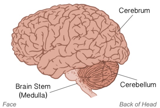
Figure 1: Major parts of the brain (Image taken from bio.libretexts.org)
Just as you begin to wonder how one organ, which is practically the size of two fists, can carry out such a diverse variety of actions, studies begin to unravel previously unrecognized functions of the brain. The gut-brain axis is one such exciting new area wherein the brain “talks” to the gut and vice versa thereby influencing the functioning of each other. Another prominent and extremely astonishing function that has recently emerged is the ability of the brain to control immune responses by modulating inflammatory signals. In this context, Columbia postdocs Hao Jin, Mengtong Li and their colleagues have very beautifully uncovered exactly how the brain serves to “sense” inflammatory cues and send signals to the body to respond correctly to these cues in their pioneering work published in Nature.
Inflammation is an immune response. It is of paramount importance to have an equilibrium between pro-inflammatory (which perpetuates inflammation) and anti-inflammatory (which curtails inflammatory signaling pathways) states in our systems to enable a potent defense mechanism. Both hyperactivation and hypoactivation of these responses can have dire consequences in organismal physiology. So how does the body-brain axis orchestrate this inflammatory balance?
Jin et al., have shown how the brain plays a significant role in determining this balance thus contributing to immune homeostasis. They have very carefully dissected the body-brain circuit controlling this communication between the immune system and brain. They have identified the neuronal populations which are activated by an incoming immune insult and how these then trigger and direct the balance between pro-inflammatory and anti-inflammatory responses.
Briefly, a peripheral immune insult is sensed by vagal neurons (part of parasympathetic system) which then transmit the signal to a distinct region of the brain, caudal nucleus of the solitary tract (cNST) in the brainstem region. When cNST neurons were inhibited, there occurred a profound elevation in pro-inflammatory response and a corresponding decrease in anti-inflammatory response. This ultimately causes the immune regulation to go haywire. On the contrary, when these cNST neurons were activated, anti-inflammatory responses were upregulated whereas pro-inflammatory responses were downregulated (Figure 2).
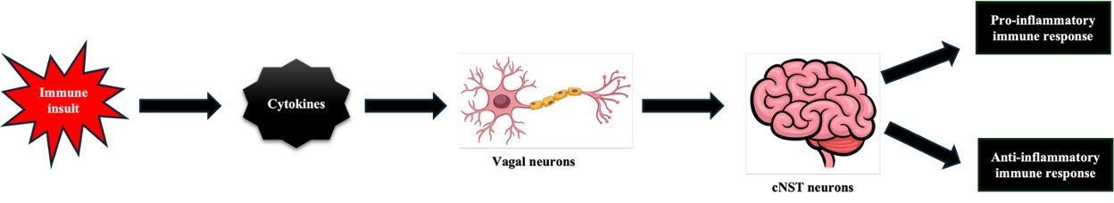
Figure 2: Schematic representation of components of the body-brain axis orchestrating inflammatory states and organismal immune homeostasis (Images of neuron and brain taken from Adobe Stock and iheartcraftythings.com).
The study also shows distinct neuronal clusters in cNST which respond to the immune response. It was further shown how cytokines, which are signaling molecules released from immune cells downstream in response to an upstream peripheral immune insult, signal specifically to vagal neurons which then propagate the signal to cNST neurons to perpetuate the inflammatory response. Vagal neurons also have specific lines of signalling, one distinct population of neurons carry anti-inflammatory signals whereas another set carry proinflammatory signals to cNST neurons.
Thus, this study uncovers a remarkable crosstalk between the brain and the rest of the body which serves to crucially maintain immune homeostasis. Modulation of intricate components of this axis holds therapeutic potential in either selectively impeding a heightened pro-inflammatory immune response or activating an anti-inflammatory state to alleviate a dysregulated immune state.
Reviewed by: Erin Cullen, Margarita T Angelova