Ever wonder how your brain directs your attention to certain stimuli or events? For example, if you are walking into a crowded restaurant you will likely direct your attention to finding an empty seat or finding your friends rather than the color of the tablecloths or the number of tables and chairs in the restaurant. Our brains have to navigate and distinguish what is important in complex environments. The term “salience” is defined as a noticeable or important object that stands out from the surroundings or background. Salience processing has been studied in neuroscience and psychology in order to understand how our brains distinguish important stimuli. This phenomenon involves two general mechanisms: bottom-up processing in which sensory information can be amplified or filtered, or top-down processing which focuses on goal-directed behaviors and cognitive control. Prior research has identified areas of the brain that are involved in salience processing using functional magnetic resonance imaging (fMRI) studies. FMRI is a technique that measures brain activity by measuring minute changes in blood flow. The regions identified include the regions in the cortex: dorsal attention network (DAN), salience network (SN), sensory cortex, primary somatosensory cortex (S1), and subcortex (Figure 1). However, understanding how these regions communicate with one another in salience processing is still unknown. In addition to these cortical networks, the locus coeruleus (LC), which is the primary source of the neurotransmitter norepinephrine (NE) and located in the brain stem (Figure 1), is also associated with salience processing.
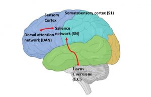
Figure 1. Cortical and non-cortical regions of the brain involved in salience processing.
Researchers commonly use MRI or FMRI (functional magnetic resonance imaging) as a technique to detect active areas of the brain while performing an activity. This is complemented with EEG (electroencephalogram) scans to measure electrical activity in the brain while performing different tasks. One of the widely used experimental designs to evaluate salience processing includes the oddball task. For these experiments, subjects are instructed to detect infrequent target stimuli (a sound of a laser gun) in a stream of standard stimuli (standard tones). FMRI and EEG measurements were obtained to determine activation of the different areas of the brain while hearing the sound of the target laser gun as compared to hearing the consistent standard tone. Columbia postdoc Linbi Hong and colleagues in their study, published in PLOS Computational Biology on May 2023, wanted to further understand the interaction between the non-cortical, LC-NE system and the cortical networks using simultaneous recordings of pupillometry which measures changes in pupil diameter and has been used as a marker for LC activity, electroencephalography (EEG) and fMRI in an oddball experiment.
The study first utilized EEG-informed fMRI analysis to map the neural cascade underlying salience processing and identify the order of specific regions which are activated during the oddball task. The researchers defined areas in the brain to understand the organization of regions involved in salience processing. Next, the researchers wanted to understand the functional connectivity between the distinct cortex regions and the LC-complex (non-cortex regions) during salience processing by analyzing EEG signals at different times during the oddball experiment. It was determined that the prefrontal cortex and dorsal attention network are the main players in processing salient stimuli.
Next, the researchers characterized the directional interactions between the functional network regions including the cortical and non-cortical regions. Specifically, they determined that non-cortical regions showed significant functional connectivity with dorsal auditory attention networks and salience networks. These results indicate that the non-cortical regions (LC network) are involved in switching between the different cortical networks during salience processing.
Overall, this study identified spatiotemporally the connectivity of different cortical regions to the non-cortical regions (LC complex) in salience processing. The study also provides insights into utilizing noninvasive pupillary response, which measures non-cortical region (LC) activity, in combination with EEG and fMRI signals, which measure cortical activity, as a valid approach to understand connectivity within the different regions in the brain. As a result, these methods could be used to determine any decline in connectivity of the cortical and non-cortical regions which can provide further understanding in neurological diseases such as Alzhiemers in which the LC network is the first region affected. Future studies from this group can provide additional input to how our brains can distinguish important information such as finding your friends in a crowded restaurant and whether this information is captured the same way in diseased conditions. So, next time you look around in a crowded restaurant, you can think, what captures your attention?
Reviewed by: Trang Nguyen, Martina Proietti Onori, Maaike Schilperoort