Neural mechanisms of Camouflage: insights from Dwarf Cuttlefish atlas

Camouflage is a common and essential survival strategy that is widely shared across the animal kingdom from reptiles, amphibians and fish, to birds and even some mammals. This ability allows them to hide in plain sight, but it is also a useful tool for hunting and a means of communication. Have you ever wondered how some animals can camouflage?

Fig1: Adult dwarf cuttlefish ( 8cm in total body length)

The dwarf cuttlefish (Fig.1) is renowned for its rapid and complex camouflage abilities, controlled by its brain. Five pairs of nerves emerge from the different brain regions and project to the different parts of the body (mantle, arms and fin) to regulate its skin’s chromatophores (these specialized cells contain different pigments, such as black, red, yellow, or brown, which contribute to the overall coloration of the skin). 

Even though we usually crown the octopus as the master of disguise, the cuttlefish is, in fact, at the top of this game. The reason for this is that the cuttlefish’s skin possesses more color changing chromatophores per square inch than its fellow cephalopods (up to 200 per square millimeter to be precise). Recently, Columbia postdoc Tessa Montague and her colleagues introduced a groundbreaking brain atlas for the dwarf cuttlefish, *Sepia bandensis*. The team used a combination of Magnetic Resonance Imaging (MRI), deep learning, and histology to create this comprehensive neuroanatomical roadmap, which is now available online through the interactive web tool Cuttlebase (https://www.cuttlebase.org/). This study aims to understand how the cuttlefish brain processes visual information to achieve camouflage. Interestingly, despite that the brain has a global volume close to the one of a mouse (94% similarity), its organization is widely different. For example, the optic lobes that only represent a few percent in the mouse brain comprise 75% of the brain in the cuttlefish, indicating their crucial role in visual processing and behavior related to camouflage and environmental interaction. The research revealed significant insights into the neural basis of cuttlefish behavior and offered a valuable resource for further studies on neural representation and brain function. 
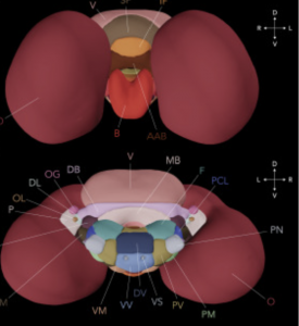
Fig. 2: Frontal (top) and posterior (bottom) 3D views of cuttlefish’s brain (from Cuttlebase)

To achieve this, the team scanned the brains of eight cuttlefish using MRI. In combining manual segmentation with deep learning techniques to extract each brain from its surrounding tissue, they created the first merged, annotated template brain (Figure  2). 

With such  3D model of brains like Cuttlebase, scientists can target brain regions to investigate precisely the function in the camouflage behavior. 

The 3D and histological brain atlases presented here are invaluable tools for studying how cephalopods, like the dwarf cuttlefish, control their behavior through their brains. 

With the help of the interactive platform Cuttlebase, researchers can precisely guide instruments for brain experiments and identify specific brain regions for their studies. These atlases also allow scientists to explore how genes and neural activity are distributed across different areas of the brain.

The brain atlas not only helps in understanding cephalopods but also serves as a resource for comparing the brain structures of different species. While little is known about the biology of the dwarf cuttlefish (Sepia bandensis), the common cuttlefish (Sepia officinalis) has been extensively studied to understand behavior, learning, memory, and skin pattern control. Despite the differences in their sizes, behaviors, and habitats, the data shows that their brain structures are quite similar. For example, Sepia bandensis lives in the Indo-Pacific coral reefs and uses high-frequency skin patterns to blend into its tropical environment. On the other hand, Sepia officinalis lives in the rocky waters of Europe and uses larger patterns to resemble rocks. Both species display unique aggression patterns during social interactions: Sepia officinalis shows a zebra pattern, while Sepia bandensis has a stippled pattern. These differences in behavior might be better understood through studies of neural activity.

This atlas is also useful for understanding evolutionary processes by comparing species that are evolutionary distant but still share similar anatomical organization. Thus, a recent study shows that despite about 600 million years of evolution, cuttlefish brains share some features with those of fruit flies. For instance, both have a central brain with two large optic lobes on the sides and learning and memory centers. Recent studies even suggest molecular similarities between certain brain cells in octopuses and fruit flies, highlighting the evolutionary connections.

This comprehensive brain atlas, combined with molecular and cellular maps, offers a deeper understanding of how these intelligent invertebrates evolved their complex cognitive abilities and behaviors. Comparing this atlas to those of other species, like the nematode C. elegans or the Human Connectome Project, provides valuable insights into brain function across different phyla. Such comparisons are crucial for uncovering universal principles of neural organization and function as well as the distinct evolutionary paths and ecological requirements of mammals and cephalopods reflected by neuroanatomical differences.

The creation of detailed brain atlases is foundational for advancing our knowledge of brain anatomy and behavior across the animal kingdom. These studies highlight the critical role of brain atlases in exploring neural structures and functions, significantly contributing to neuroscience. Despite the existing atlases, there is a pressing need for more to fully understand brain evolution throughout the animal kingdom.

By expanding the collection of brain atlases across a wider range of species, researchers can uncover evolutionary pathways and adaptations that have shaped cognitive abilities. The detailed information provided by these atlases helps correlate brain structure with function and behavior, offering insights into the neural basis of complex traits. Increasing the number of brain atlases will enable scientists to trace the development of neural mechanisms from simple organisms to complex beings, enhancing our understanding of brain evolution and capabilities.

Investing in the creation of more brain atlases will build a robust framework for comparative neuroanatomy, deepening our understanding of individual species and illuminating broader evolutionary trends. The insights from these comparative studies are crucial for developing new hypotheses about brain function, improving neural models, and advancing our knowledge in neuroscience and related fields. If you want to know more details check out the original paper.

Written by: Nathalie Houssin

Reviewed by: Trang Nguyen,  Margarita Angelova, Jerry Gourdin

Layer by layer – How reducing the thickness of layered magnetic materials can change tomorrows electronics

By Daniel Čavlović


What if I told you that nearly all electronic devices like phones or computers waste incredible potential in the way they are designed to work? All currently used electronic devices move electrons from one place to another creating a current relying solely on the particle’s negative charge. Very old-school physics known for centuries. But electrons are more than just charges! According to quantum mechanics – yes bear with me – an electron always possesses an additional intrinsic property, its spin, which can be either spin-up or spin-down (often depicted with arrows). These spins are slowly moving into the attention of applied research even though the quantum mechanics behind it is known for one century. The next-generation of electronics will leverage the full potential of the disregarded information stored in spins. But to build a so-called spin transport electronic (spintronic) device we need to find materials that are able to store, transport, and switch the spin information.

A group of researchers has synthesized and studied a novel material TaFe1.14Te3 which could be perfect for spintronic applications. By pressing elemental tantalum (Ta), iron (Fe), tellurium (Te), and some tellurium chloride into a pellet, sealing in a fused glass tube under vacuum and heating between 600 – 700 °C for a week they obtained needle-like crystals of this material. What makes this newly forged material so special is the way of how the elements are connected to each other. Even though the crystals resemble three dimensional needles, the underlying fundamental structure shows stacked layers of two dimensional sheets. Just like a stack of papers, each sheet is firmly connected and requires scissors to cut the sheet. On the other hand only loose forces keep the stack of sheets together and as little as a bit of wind blows the stack of paper away). Amusingly, the same way that you can remove the top sheet from a paper stack with tape, this quite literally works the same way with TaFe1.14Te3 (see Figure 1 below). This Nobel Prize winning technique is known as mechanical exfoliation and led to the isolation of the first in-depth studied 2D material, graphene. Graphene is a single layer of graphite (mistakenly called ‘lead’ in pencils). In graphite each layer of graphene interacts with the layers in between it’s sandwiched in. Thus, all defects or inconsistencies are diffused into and covered up by the mountain of layers. Like picking out the best singer from the choir for a solo, by reducing the number of layers of the material the properties start to change and become more featured and outstanding. In the case of graphite (the pen material) one single layer of it—graphene—becomes incredibly conductive and mechanically robust. Cool properties but not very useful for electronic devices because graphene has no band gap. It’s pretty much just a “wire” which always conducts electricity. In contrast a band gap is like a wire with a switch, where two states can be addressed (for binary information). Single layers of TaFe1.14Te3 indeed do possess a band gap and are magnetic too, both properties required for spintronics. Its magnetism is especially interesting: The studies revealed that in each single layer of this material there are quasi-one dimensional chains of iron atoms decorated with unpaired electrons whose spins give rise to its magnetism. In a single layer all neighboring spins align and point in the same direction, a property called ferromagnetism or 2D magnet.

Figure 1: Schematic side-view of a TaFe1.14Te3 layer being pulled off as an illustration of mechanical exfoliation.

You can imagine each of those spins in iron atoms like a compass needle always aligning with the earth’s magnetic field. But what happens if you stack another layer on top of it? The top layer’s spins align in the opposite direction with respect to the layer below forming an antiferromagnetic bilayer. This shows that the spins between two layers strongly interact with each other. While a single layer interacts strongly with an external magnetic field from let’s say a magnet closeby the bilayer does not care about external magnetic fields because of the much stronger interaction with its neighboring layer. You may have heard that a magnet can damage or wipe your computer’s hard drive, which is true for older technology which relied on ferromagnetic layers. One elegant solution to this is to store information in antiferromagnetic bilayers which further enables minimizing the size and amount of materials needed for electronic devices.

The researchers have studied TaFe1.14Te3 and its electronic structure in even more detail. They found that the material shows metallic behavior, further supported by calculations, and itinerant magnetism but at the same time local moments which makes the exact electronic structure complicated to determine. What this means is that as a whole it shows local sites where the magnetism arises from but is spread out and averaged as it never resides fully localized. The underlying origin of this special magnetism is still subject of further studies of this material which is probably being deciphered at this very moment.

The most important property–stability under ambient conditions–may sound boring but is essential for practicable applications. Even though there are other 2D materials known which show similar remarkable properties but their translation from fundamental research towards application is not really feasible because they react with trace water or oxygen from the air we breathe!

Even though spintronic devices are far from being commercialized, one day you might hold a superior and more energy efficient spintronic phone in hand and admire the success of technological advancement. Remember, that all originated from fundamental research which managed to leverage the hitherto elusive spin information with 2D materials like TaFe1.14Te3


Original paper

Reviewed by: Trang Nguyen


Unlocking the code of longevity: How your diet might hold the key to a sharper mind

By Divya Vimal

We’ve all grown up hearing about the virtues of eating our greens and maintaining a healthy diet. But what if I told you that beyond just keeping your body in shape, a healthy diet could be your secret weapon against the ravages of aging and even dementia in later life? Yes, that’s right- those veggies might just be your ticket to a sharper mind and a healthier future! A groundbreaking study delved into this intriguing possibility, focusing on whether a healthy diet could slow down the biological clock and reduce the risk of dementia.

The researchers examined information from the Framingham Offspring Cohort, consisting of 1,644 participants aged 60 years and older, and has been under follow-up since 1971. They used algorithms also called epigenetic clocks to measure how quickly participants were aging biologically by assessing the methylation status of the DNA in white blood cells. One such epigenetic clock is DunedinPACE, which was utilized in this study as a predictor of health-span and lifespan. Meanwhile, they assessed diet quality using the Mediterranean Dash Intervention for Neurodegenerative Delay diet (MIND) diet score and tracked the occurrence of dementia and mortality. The MIND diet has been developed for prevention of dementia, combining key principles from two healthy diets (i.e. Mediterranean diet and Dietary Approaches to Stop Hypertension Diet). The MIND diet emphasizes high intake of neuroprotective foods such as fish, green leafy vegetables, berries, and nuts, while minimizing intake of red meat, butter, sweets. The MIND diet is a composite scoring system based on components of the MIND diet. Each component of the diet was scored from zero to one, reflecting the frequency of food consumption. Scores were assessed weekly, and the total MIND diet score, ranging from 0 to 15, was calculated as the sum of the 15 component scores. Higher scores indicated better adherence to the diet over the long term. 

Excitingly, the results revealed a compelling link between diet, aging, and dementia risk. Participants who adhered more closely to the MIND diet enjoyed a slower pace of biological aging (each 1-standard deviation (SD) increase in MIND diet score associated with a 0.20-SD slower DunedinPACE). For each improvement in the MIND diet score, there was a reduction of 34 fewer incident dementia cases per 10,000 people a year, suggesting that a healthier diet could lead to fewer cases of dementia over time- a striking finding! Digging deeper, about 27% of the diet’s impact on dementia risk was attributed to its effect on the aging pace. This suggests that a good diet might protect your mind by keeping your body younger at the cellular level.

Furthermore, a slower pace of biological aging was independently associated with lower risks of both dementia and mortality. This means that not only does a healthy diet potentially safeguard your mind, but it could also help you live longer.

Covariate analysis (statistical method) including factors like socio-economic status, genetics, lifestyle, and health conditions didn’t change the main results. Interestingly, there were no significant differences in how diet affected aging based on sex or APOE4 status (a genetic risk factor for Alzheimer’s). Furthermore, the study revealed that maintaining a healthy diet at different life stages, from mid-life to older age, was associated with slower biological aging, reduced risk of dementia, and lower mortality rates. Alternatively, similar analysis with the Mediterranean Diet Score and the Dietary Guideline Adherence Index mirrored the associations between different measures of diet quality and health outcomes underscoring the well-known health benefits of the Mediterranean diet. These findings collectively underscore the significant impact of diet on aging and health outcomes, emphasizing the importance of maintaining a healthy diet throughout life.

While the study had its limitations, such as a predominantly white cohort and participant recall bias, its implications are profound. It hints at the possibility that by monitoring our biological age, we might gain valuable insights into how to prevent dementia and age-related decline. Furthermore, the study couldn’t differentiate if a healthy diet directly affected organ health, leading to slower aging (egg before the chicken) versus its impact on cellular aging indirectly preserving organ health (chicken before the egg).

In essence, this study highlights the power of diet in preserving both body and mind. It paints a hopeful picture of a future where something as simple as dietary choices could be the key to a longer, healthier life with a more agile mind. 

Original paper

Reviewed by: Aline Thomas and Trang Nguyen

Beyond Diabetes: Exploring Metformin’s Promise in Prostate Cancer Therapy

By Rafaela Muniz de Queiroz 

Cancer and diabetes are two major contributors to mortality in patients, ranking among the top 10 causes of death worldwide by the World Health Organization (WHO). Both diseases pose a significant challenge to public health systems, representing a complex challenge that requires comprehensive strategies to reduce their impact on global mortality rates. Metformin, among other drugs, is the main first-line prescribed medication for the management of type 2 diabetes, and it is known for its efficacy and tolerability. Yet, metformin has gained attention for its role in other contexts, including cancer and cardiovascular disease.

Prostate cancer is a significant global health concern and a leading cause of cancer-related deaths among men. While treatments like radical prostatectomy (RP) and radiotherapy are effective for many patients, for others, the disease progresses leading to metastasis, which can be difficult to treat and is often fatal. Previous studies have pointed to metformin’s potential role in prostate cancer, but conflicting findings have led to an inconclusive verdict on the use of metformin for this disease. A recent breakthrough study headed by Alexandros Papachristodoulou from Dr. Abate-Shen’s group in the department of Molecular Pharmacology and Therapeutics at Columbia University Irving Medical Center, has shed light on a potential new avenue for the use of an anti-diabetic drug in the treatment of prostate cancer.

The prostate gland is susceptible to inflammation and oxidative stress, both of which can accelerate prostate cancer progression. Interestingly, oxidative stress influences the behavior of NKX3.1, a gene that plays a crucial role in protecting the prostate epithelium from cancer-related stress. In their work, Dr. Papachristodoulou and colleagues examine the interplay between NKX3.1 and the widely used diabetes drug metformin. The interplay between NKX3.1 and metformin are found in the mitochondria. NKX3.1 helps protect against harmful free radicals (unstable molecules that can damage cells by stealing electrons from other molecules) and supports normal mitochondrial function, while metformin has a known ability to regulate mitochondrial function and reduce oxidative stress.

The study published in 2024 used a comprehensive number of strategies to investigate its hypothesis, including in vitro work with the use of human prostate cancer cell lines, in vivo work using mouse models, and analysis of retrospective cohorts of patients. The authors show an impressive reduction of tumor size in mice under oxidative stress, by exposure to the herbicide Paraquat, and treated with Metformin. This difference was only seen if tumors did not express NKX3.1. They show that metformin treatment can fully rescue mitochondrial function that had been lost upon oxidative stress in prostate cancer cells lacking NKX3.1, pointing to a possible molecular mechanism for the effects seen in mice.

Moreover, the study analyzed the biochemical recurrence (BCR)-free survival, evaluated by the blood levels of PSA, the antigen used to diagnose and surveil prostate cancer in patients. This parameter is used in the clinic to show the percentage of patients that remain disease-free after RP over time. Utilizing data from two cohorts of patients, the researchers showed that those expressing low levels of NKX3.1 and taking metformin had a much higher rate of BCR-free survival compared to patients not taking the medication. The group of patients expressing high levels of NKX3.1 showed no difference in BCR-free survival when taking or not the drug. In addition, when the disease progression of a group of prostate cancer patients that have been followed up for up to 10 years was investigated, the authors observed that among the patients with low-NKX3.1, all patients treated with metformin evolved to have their disease classified more favorably during follow-up, while most patients not exposed to the medication had a worse development of the disease.

These findings open exciting possibilities for personalized treatment strategies for prostate cancer. It offers hope for a future where precision medicine plays a pivotal role in combating this disease and improving patient outcomes. By identifying patients with low NKX3.1 expression levels, clinicians may be able to tailor metformin therapy to those who are most likely to benefit, potentially improving patients’ quality of life and extending survival rates. While further research and clinical trials are needed to validate these findings, this study represents a significant step forward in understanding the biology of prostate cancer and exploring novel therapeutic avenues. 

Original paper

Reviewed by: Trang Nguyen, Erin Cullen

Who brings the SARS-CoV-2 virus home and who spreads it around?

By Arpit C Swain

A child, a teenager and an adult enter a household… No, this is not the beginning of a joke. This is the beginning of a question for you. So, a child, a teenager and an adult enter a household, who do you think would be most likely to infect others in the household? If I were to venture a guess, I would say the child would be most likely to infect others in that household. The child has the most underdeveloped immune system among the three after all, and I have frequently heard the ‘must have been because of the daycare’ rhetoric from young parents who fall sick during the flu season. However, I would be wrong, at least if it’s the SARS-CoV-2 virus.

In their recent article, Christiaan H. van Dorp, a postdoctoral researcher at Columbia University Medical Center, and his colleagues find that adults are most likely to introduce an infection into a household, closely followed by teenagers, whereas children were only ~60% as likely as adults to introduce an infection into a household. They estimated that the rate at which an adult introduced an infection into a household was 0.0008 per day in mid-October 2020, which means that a group of 1200 adults infect one uninfected person every day.

The authors of this article used a prospective household study rather than a reactive household study. A reactive household study is not suitable to estimate how often an infection is introduced into a household as only households that are already infected are included in the study and the time of the infection is unknown. A prospective household study, in contrast, follows all participating households semi-passively during the at-risk period. In the Dutch study that Christiaan and team used for their analysis, more intensive follow-ups were performed upon notification of acute respiratory symptoms in the household.

The participating households were tracked for a maximum of 161 days. The team found that SARS-CoV-2 was introduced and established in 59 out of 307 households (~19%) between August 2020 and March 2021. The surge in hospitalization of hundreds of people in the same period closely followed the number of household infections (Figure 1). The peaks in hospital admissions (see the yellow dots) were always 1-2 weeks after the peak in the household infection rate.

Figure 1: The number of infections in the households is closely followed by the number of hospitalizations reported. The blue line is a measure of the infection rate of a household and the gray region around it is the variation in that rate.

The Netherlands was under lockdown during the period of the prospective household study. Christiaan and his colleagues’ analysis did not test whether children were less likely to introduce the infection into a household because of their limited interaction with the outside world during the lockdown. However, they tested whether the age of a person was a factor in how fast the virus was transmitted within a household upon its introduction into the household. They categorized the population into three different age groups: children (0-12 years old), adolescents (12-18 years old) and adults (above 18 years old). The team formulated a ‘full within household model’ where they tested all possible interactions between the three different age groups (see Figure 2). They found that once an infection had been introduced into a household, children were the primary age group that transmitted the infection within the household. The transmission rates due to adolescents and adults within a household were at most half of that of children, the rates for adolescents being marginally higher than that for adults (Figure 2).

Figure 2: All possibilities of transmission among the three age groups were tested in the full within household transmission model.

Using the full within household model again, Michiel van Boven, a researcher at University Medical Center Utrecht in the Netherlands, tested the effectiveness of a vaccine in controlling the spread of infection within a household. He assumed that a vaccine is ‘leaky’, which means that a vaccinated individual who contracts the virus would not be infected, however, can still spread the virus to others in the household. The simulations showed that vaccinating just the adults was effective in controlling the infection in the household. Vaccinating the adolescents, on top of the vaccination of adults, did not improve the control of infection. It is surprising that although adolescents are likely to introduce the infection into a household and also transmit it within the household, their vaccination does not improve the control of infection.

Christiaan and the team’s results from the prospective household study, therefore, shows that an adult brings the SARS-CoV-2 virus home which is then primarily transmitted within the household by children. Perhaps I was partially correct. A child would be most likely to infect the household, if it is already infected, otherwise it’s the adult who is most likely to infect the household.

Reviewed by: Christiaan van Dorp, Erin Cullen, Trang Nguyen, and Giulia Mezzadri


Shining a Light on Microscale Innovations

What comes to mind when you think of light? Perhaps the vision of yourself basking in the sun on a California beach. Or the most recent book of Michelle Obama “The Light We Carry”. Or even the metaphorical light we’re not supposed to follow when our time comes. Here, we are going to talk about a concept even more fundamental: the physics of light.

Light, with its dual nature as both a wave and particles known as photons, exists in different “forms”, each characterized by its unique wavelength or color. For instance, the wavelength of the visible light, which is the electromagnetic radiation detectable by the human eye, ranges from 400 to 700 nanometers.

Image credits: Chiara Trovatello

Fascinatingly, we can manipulate light and change its wavelength using a powerful tool: nonlinear optics. Inside nonlinear materials light behaves in a “nonlinear” manner, meaning that its response is not directly proportional to the input. In such systems, the properties of light change as it interacts with the material, resulting in nonlinear phenomena. For example, photons interacting with a nonlinear material can be transformed, with a certain probability, into new photons with twice the frequency of the initial photons in a process called Second Harmonic Generation (SHG). Another phenomenon, even more intriguing, is the Spontaneous Parametric Down-Conversion (SPDC), in which one photon of higher energy is converted into a pair of entangled photons of lower energy. What makes these entangled photons unique is their intrinsic interconnection, even when they are separated by vast distances, or even placed at opposite corners of the universe. Changing the state of one of them will result in an instant change in the state of the other photon.

Nowadays, SPDC is especially relevant since it’s at the heart of many applications, for instance in quantum cryptography for secure communications and also for testing fundamental laws of physics in quantum mechanics. Inducing these nonlinear behaviors requires crystals with large thickness, typically of the order of a few millimeters. This requirement is due to the relatively low nonlinearity of these materials. Indeed, when the nonlinearity of a material is low, a larger volume of material is required in order to achieve an equivalent level of efficiency. This limitation has been overcome thanks to the innovative work of Chiara Trovatello, a Columbia postdoc from the Schuck Lab. She and her colleagues have successfully developed a method for generating nonlinear processes, such as SHG and SPDC, in highly nonlinear layered materials with remarkable efficiency, down to sizes as small as 1 micrometer – a thousand times thinner than a millimeter! To put this in perspective, the materials that Chiara and her colleagues have realized are 10-100 times thinner than standard nonlinear materials with similar performances.

While materials with a high conversion efficiency were already well-known in the field, achieving macroscopic efficiencies over microscopic thicknesses was still an open challenge. The critical component was to find a way to achieve the so-called phase matching condition in microscopic crystals with superior nonlinearity. Phase matching ensures efficient conversion of light by maintaining the fields of the interacting light waves in-phase during their propagation inside the nonlinear crystal. To this end, Chiara and her colleagues designed and realized multiple layers of crystalline sheets, stacked with alternating dipole orientation. With this procedure, they were able to change the way the material responds to light, meeting the phase matching condition over microscopic thicknesses. This methodology provides high efficiency in converting light at the nanoscale, and unlocks the possibility of embedding novel miniaturized entangled-photon sources on-chip. It paves the way for novel technologies and more compact optical devices, for applications in quantum optics. Projecting ourselves into the future, it’s not hard to envision a world where everyone will carry a sophisticated quantum device in their pockets, this time embracing the physical light rather than running away from it.

Reviewed by: Trang Nguyen

Unlocking the Mysteries of Muscle Contraction: A Breakthrough in Ryanodine Receptor Structure

Ever wonder how our muscles work when we move? It all comes down to calcium—a key player, released from storage spaces within our muscle cells called the sarcoplasmic reticulum (SR), that regulates the muscle contraction. The crucial protein allowing calcium release is the type 1 ryanodine receptor (RyR1), an ion channel of enormous proportions (the largest known so far) that is embedded in the SR membrane.
RyR is more than just a protein; it’s a critical piece of the puzzle in understanding diseases like muscular dystrophy and heart failure. Plus, RyR serves as a potential pharmacological target, opening avenues for therapeutic interventions in muscle-related disorders.
Yet, working with such bulky proteins in its native state – within the cell membrane- is a challenging subject. Typically, the steps needed to resolve the structure of proteins normally involve using detergents and harsh conditions that disrupt the native state of a protein.
However, why is it crucial to comprehend the structure of proteins in their native state?? Because it enables us to mimic the conditions in living cells and gain valuable insights into their physiological relevance, their interaction with other molecules, exploring disease mechanisms, and facilitating drug development, among others.
So far, it has been used cryogenic electron microscopy (cryo-EM), an imaging technique that freezing samples and using electrons instead of light to reveal the protein’s intricate 3D structure (i.e., protein’s shape, how its various parts are arranged, and the interactions between its amino acids). While the 3D arrangement of the RyR1 protein had been solved at a very detailed level (referred to as the “High-resolution structure”), the configuration in its native state was still unknown.
In the pursuit to unravel the structure of proteins within native or near-native membranes, a recent breakthrough has provided a high-resolution view of RyR1.
Departing from traditional detergent-based methods used to solubilize and stabilize membrane proteins, Dr Melville Zephan, CUIMC postdoc, and colleagues adopted a gel-filtration approach with on-column detergent removal, they’ve captured RyR1 within liposomes—tiny bubbles mimicking cell membranes (Figure 1).

Figure1. Flow diagram illustrating protein-incorporated liposomes.
RyR1 from rabbit skeletal muscle was homogenized and purified using ion-exchange chromatography, a technique that separates compounds based on their electric charge. Liposomes, which are tiny vesicles made of self-assembly of lipid bilayers in water, were formed using gel filtration. This process helped incorporate RyR1 into liposomes and remove any excess or empty liposomes. (Adapted from Melville Z. Cell Press 2021)

This novel technique maintains a more natural environment, allows to purify membrane proteins and remove detergent molecules in excess, offering a clearer glimpse into how RyR1 functions (Figure 2).

Figure2. RyR1 representation in native and near-native membranes incorporated in liposome after gel filtration. (Adapted from Melville Z. Cell Press 2021)

Their study reveals how RyR1 behaves in its natural environment, forming a network of channels within cell membranes. While their investigation in liposomes gives only a partial view of this complexity (without seeing how molecules interact), it sets the stage for future investigations. This approach, extending beyond RyR, offers a blueprint for studying membrane proteins in native conditions.

In summary, this work successfully captured the intricate structure of RyR1 in liposomes opening doors to a better understanding of muscle physiology and offers a sharper lens for exploring the complex world of membrane proteins.

For more details on this research, refer to the article: “High-resolution structure of the membrane-embedded skeletal muscle ryanodine receptor”.

https://www.sciencedirect.com/science/article/pii/S0969212621002963?via%3Dihub

Reviewed by: Trang Nguyen, Carlos Diaz, Erin Cullen


“super-SOX” hits iPSC research out of the park

The human body contains trillions of cells, which have extremely diverse functions. Remarkably, every one of these cells has the same genetic code. This diversity is possible because of the developmental process of differentiation. As an organism grows, cells take on traits that are useful in a particular context. For example, skin cells produce a protein called keratin that makes skin tough in order to protect the rest of the body from the outside world. Other cells don’t produce keratin because it would hinder their function. Once cells have differentiated and taken on a particular role, they can only generate more of the same kind of cell – skin cells only divide into more skin cells. This is because sections of DNA are hidden away during differentiation and stay hidden after the cell divides, ensuring that all the cells in a particular organ (like the skin) act like they’re supposed to.

To turn one type of differentiated cell into another was once thought to be impossible. The only cells that can generate any type of cell in the body are pluripotent stem cells (PSCs). PSCs have many potential applications in science, medicine, and industry, including disease modeling and drug testing as well as cell-based therapies and biotechnology. The opportunity to study PSCs can also enable scientists to answer basic questions about mammalian development. 

In 2006, Japanese researchers Takahashi and Yamanaka discovered that PSCs actually can be created from differentiated cells. Doing so requires just four key proteins: Oct4, Sox-2, Klf4, and cMyc. These proteins together are referred to as the Yamanaka factors. The discovery that the Yamanaka factors could turn a differentiated cell into a PSC was incredibly exciting to the scientific community, earning Dr. Shinya Yamanaka and a colleague the Nobel Prize in Physiology or Medicine in 2012.

Although this discovery created a broad array of new possibilities in science, it turns out that making PSCs in a laboratory is tough. Making healthy, high-quality PSCs from different species is even tougher. Induced pluripotent stem cells (iPSCs) have only been generated from a few species, and some cell lines create healthier iPSCs than others. A clearer understanding of how the Yamanaka factors interact with DNA and each other could reveal ways to make iPSC generation more robust and consistent, and realize the exciting potential of iPSCs. 

This challenge was taken on by a research group led by Drs. Sergiy Velychko and Caitlin MacCarthy in collaboration with CUIMC postdoc Dr. Vikas Malik and other teammates around the world. The group published a study this month in Cell Stem Cell describing a new protein based on the Yamanaka factor Sox2. They aptly call this protein “super-SOX” based on a number of studies demonstrating its ability to better generate iPSCs. The experiments also uncovered basic principles of pluripotency and development.

While three of the Yamanaka factors can be replaced with other proteins from the same family to generate iPSCs, Oct4 is the only member of its family (the POU family) that can induce pluripotency. The researchers set out to understand this oddity, with the goal of revealing the molecular interactions that lead to pluripotency. They first found that while a different member of the POU family could not generate iPSCs with the Yamanaka factor Sox2, it could generate them with a mutant protein based on another member of the SOX family, Sox17. 

Next, the researchers created a library to test different Sox2-Sox17 chimeras – mutant proteins that include components from both Sox2 and Sox17. Amino acids are the most basic building blocks of proteins, and surprisingly, the researchers found that a chimera with only one different amino acid from Sox2, Sox2AV, stabilizes the interaction between Sox2 and Oct4 and increases DNA binding efficiency. Oct4 is distinguishable from other members of the POU family by a negative charge in its “linker” domain. This negative charge allows Oct4 to form “salt bridges” connecting it to Sox2. Swapping the Sox2 amino acid alanine for the Sox17 amino acid valine promotes the formation of these bridges, encouraging Sox2AV and Oct4 to interact and bind to DNA together. The stronger connection between these two proteins dramatically improved pluripotency. 

Photo of healthy adult mice generated entirely from iPSCs and schematic of how outcomes change dramatically after replacing Sox2 with Sox2AV (Adapted from McCarthy et al., 2024).

The researchers hypothesized that the improvements in DNA binding they observed could promote the development of iPSCs into mature organisms. Live mice and rats have previously been generated from iPSCs, but these animals rarely survive to adulthood. Replacing Sox2 with Sox2AV – a simple change of one amino acid – when generating iPSCs dramatically increased mouse survival. 10/10 different cell lines treated with Oct4, Sox2AV, Klf4, and cMyc were able to produce live all-iPSC mouse pups, which only 3/8 cell lines treated with all the traditional Yamanaka factors were able to achieve. In the best-performing Sox2AV line, 43.3% of all-IPSC mice became healthy adults, compared to only 15.2% from the best-performing Sox2 line.

A further study found that a more complex chimera of Sox2 and Sox17 (Sox2-17) could improve iPSC generation in five different species: mice, cows, pigs, monkeys, and humans. These species were chosen for their potential to bring iPSC technologies to either biomedical research or industry – for example, iPSCs from livestock species could be key to producing lab-grown meat. Non-human primates and livestock species are less established in iPSC research, but the invention of Sox2-17, or “super-SOX”, opens many doors for future uses of iPSCs from these species. 

One barrier to creating iPSCs from human cells is age – differentiated cells from adults are highly resistant to becoming iPSCs. Strikingly, the group found that although the traditional Yamanaka factors could not produce iPSCs from skin cells of certain Parkinson’s disease patients, replacing SOX2 with the chimeric SOX2-17 could produce iPSCs from these samples.  Parkinson’s disease is characterized by the selective death of neurons in a brain region important for motor coordination called the substantia nigra, and could theoretically be treated by replacing lost neurons with iPSCs differentiated into neurons with a particular patient’s genetic code. The finding that SOX2-17 improves iPSC generation in cells from Parkinson’s disease patients demonstrates an exciting potential medical application for this discovery. 

The paper goes on to provide an explanation for these exciting findings. Prior to this study, it was known that although Oct4 is necessary for inducing pluripotency, too much Oct4 impairs iPSC generation. Free Oct4 encourages proliferation, which is actually detrimental to inducing pluripotency. Here, the researchers found that the stronger bond between Oct4 and Sox2AV or Sox2-17 lowers the proliferation rate, which improves the health of the cells that are generated. They went on to show that the bond between these two proteins is a main driver of the process of reverting a differentiated cell to a “naive” iPSC, which broadens the possibilities for the cell’s future development. 

This study makes a significant practical step toward realizing the potential of iPSCs. Simultaneously, it answers longstanding questions regarding how exactly the Yamanaka factors induce pluripotency, showing that the interaction between Oct4 and Sox2 has a central role. These findings have exciting implications for iPSC research, a field at the very forefront of science and medicine. 

Reviewed by: Trang Nguyen, Giulia Mezzadri, Carlos Diaz, Vikas Malik


Revolutionizing Cancer Immunotherapy: Unveiling Novel Biomarkers through Longitudinal High-Parameter Spectral Flow Cytometry

Immunotherapy, a groundbreaking approach to treating diseases by harnessing the body’s immune system, has emerged as a cornerstone in cancer treatment. In contrast to chemotherapy, immunotherapy offers a less toxic alternative for cancer patients. Chemotherapy indiscriminately targets both cancerous and healthy cells throughout the body, whereas immunotherapy specifically focuses on attacking cancer cells. However, the challenge lies in the rarity of responses to immunotherapeutic agents, their limited efficacy across tumor types, and the unpredictable nature of outcomes.

Tumor-infiltrating lymphocytes (TILs), immune cells present in and around tumors, signify an active immune response against cancer. The presence of TILs often correlates with improved patient outcomes, making the identification of predictive biomarkers a crucial pursuit in tumor immunology. Yet, achieving this goal remains a formidable challenge.

Key players in the immune system are white blood cells, produced in the bone marrow, patrolling the body for foreign invaders. Lymphocytes (T cells, B cells, and Natural Killer cells), neutrophils, and monocytes/macrophages constitute the most common types of immune cells.

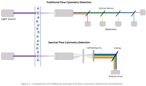
To address the need for biomarkers to guide immunotherapy, a research group led by Dr. Benjamin Izar at Columbia University Medical Center has pioneered a cutting-edge approach. They developed a 34-parameter spectral flow cytometry panel and an advanced data analysis pipeline to explore protein-level immune phenotypes across different cancer phases. Conventional flow cytometry uses dichroic mirrors and band pass filters to select specific bands of the optical spectrum for detection using point detectors. Unlike conventional flow cytometry, spectral flow cytometry captures the entire spectral profile of fluorophores, allowing for a more comprehensive understanding of immune responses (Figure 1). This innovative method enhances signal resolution by subtracting cellular autofluorescence, showcasing its potential to uncover crucial insights into cancer immunotherapy. This allows the use of more existing fluorophores that would otherwise be incompatible on a conventional flow cytometer and the expansion of immunophenotyping panels beyond 40 fluorescent parameters.

Figure 1: Comparison of traditional and spectral flow cytometry detection mechanism. Images from FluoroFinder.

In this groundbreaking study, researchers meticulously profiled various tissues affected by prostate and colorectal cancers in mice undergoing anti-PD-1 immunotherapy. Their investigation revealed a significant correlation between the expression of KLRG1, recognized as a coinhibitory receptor on T cells and NK cells, and tumor-related factors such as burden, progression, and regression in response to anti-PD-1 treatment. KLRG1 emerged as a potential marker indicative of terminal differentiation and/or senescence, influencing key cellular pathways and checkpoints.

The longitudinal high-parameter spectral flow cytometry approach employed in this research showcased its prowess in extracting novel targets and biomarkers from dynamic ‘temporal atlases’ of antitumor immunity. This innovative methodology holds the promise of unraveling the intricate, ever-changing interactions within the tumor microenvironment. By providing a deeper understanding, it aims to enhance the efficacy of immunotherapy in clinical applications. The study particularly emphasizes the need for further exploration of KLRG1+ CD4 T cell subsets as potential targets or prospective biomarkers for advancing cancer immunotherapy.

For more details on this research, refer to the article: “KLRG1 Marks Tumor-Infiltrating CD4 T Cell Subsets Associated with Tumor Progression and Immunotherapy Response.”

Written by: Trang Nguyen

Reviewed by: Erin Cullen

Take a look in the past to foresee the future: the evolution of genome engineering

Transposons, also known as mobile genetic elements, are segments of DNA capable of moving within the genome. This mobility can potentially induce mutations, as transposon “jumping” within a gene may interrupt the gene and trigger its loss. Transposons utilize their own enzymes, transposases, to move in and out of the genome.

Due to the potential harm caused by transposons, cells continually evolve strategies to control them. In response, transposons adapt, arming themselves with new strategies to evade cellular control. Mobilized transposons can hijack adjacent DNA information and transfer it to a new location, sometimes involving a gene that provides a survival advantage, thus increasing the transposon’s chances of propagation. Simultaneously, cells “steal” genes from transposons, repurposing them to function in processes advantageous to the cell. This ongoing arms race between transposons and host defense systems has facilitated a widespread exchange of genes between different life forms and is a major driver of genome evolution across all domains of life. Transposons play a crucial role in this process.

In bacteria, the CRISPR-Cas9 system is fundamental for protection against viral infections. Using a small RNA molecule called guide RNA, the CRISPR-Cas9 system acts as a nuclease, akin to DNA cutting scissors, destroying the viral genome. This system can be modified to edit genes in organisms beyond bacteria. The guide RNA is altered to direct the Cas DNA scissor to a gene of interest instead of a viral genome. This programmable gene-editing system has diverse applications, from basic biology research to disease treatments, and it earned the 2020 Nobel Prize in Chemistry for Emmanuelle Charpentier and Jennifer Doudna. Notably, scientists have recently traced the evolutionary origins of CRISPR-Cas9 to transposons.

In a recent study published in Nature, Columbia postdoc Chance Meers and his colleagues provided further insight into the evolution of the CRISPR-Cas9 nuclease system by examining how transposons use cellular nucleases to proliferate within genomes. They focused on a specific transposon system called insertion sequences (IS), which are bacterial transposons encoding only the genes necessary for transposon excision and movement to a new genomic location. This includes a transposase. However, many IS transposons contain an accessory gene that carries the information for a nuclease which functions similarly to the CRISPR-Cas system as an RNA-guided DNA cleaving enzyme.

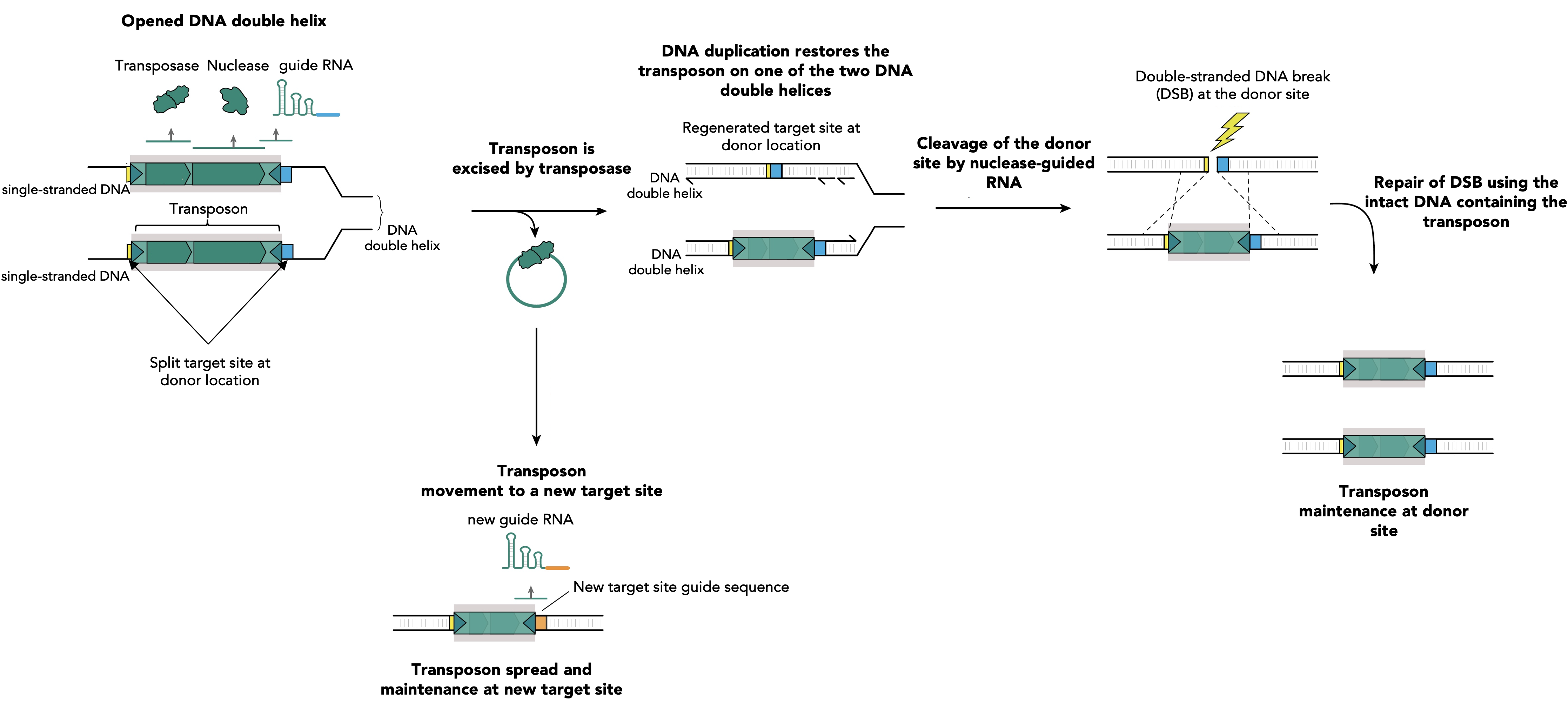
The IS element does not need a nuclease for its movement, the transposase is sufficient to mobilize the transposon to a new location. So why are so many of the IS elements carrying those RNA-guided DNA scissors? While the mechanism by which IS elements are mobilized by their transposase is similar to a cut-and-paste process, leaving no copy at the original DNA location, Meers and colleagues discovered that the rate of excision is more efficient than the rate of integration. Without an additional system for transposon proliferation, the transposon would eventually be lost since not every excised copy manages to be reintegrated. The study of the authors revealed that the accessory CRISPR-like functioning nuclease guides a copy of the IS element back to its original location, generating two copies of the element—one at the original site and one at the newly inserted site. This changes the mechanism from cut and paste to cut and copy when the transposase function is complemented by the accessory nuclease, increasing the transposon copy number present in the genome, and thus serving for the transposon selfish proliferation (Figure 1).

Figure 1. RNA-guided nucleases assist transposon survival via guiding specific breaks at donor sites and transposon restoring repair. During DNA replication the two strands of the DNA duplex are separated and each strand serves as a template for the synthesis of a new DNA molecule. Single stranded DNA facilitates the transposition mediated by the transposase of IS elements. The excision of the transposon leads to its loss at one of two DNA strands, while DNA replication restores the transposon on the other DNA strand. Simultaneously, transposon excision restores a target site at the transposon donor location which is specifically recognized by a guide RNA (blue segment). This guide RNA directs the accessory nuclease on the excision location and DNA cleavage occurs. The resulting double strand break (DSB) is lethal for the bacteria and its only chance of survival is to copy the information from the homologous newly synthesized intact DNA molecule that contains the transposon. This leads to reconstitution of the transposon at the donor site on the two newly synthesized DNA duplexes. Transposition at the new target sites will produce new guides, specific for the new insertion site location (orange segment), which will facilitate the future transposon spread and maintenance by identical mechanism. 

By developing powerful assays to track the movement of transposons within bacterial genomes, or other small DNA molecules called plasmids, as well as from one bacterium to another, Meers and his colleagues uncovered how those RNA-guided DNA cutting nucleases work. The authors’ discovery enhances our understanding of how proteins collaborate with RNA guides to target and edit genomes. In addition, the study unveils the original role of CRISPR’s nucleases from an evolutionary perspective before they were repurposed by the bacterial host genome to fight viruses, which was to serve the selfish propagation of transposons. Moreover, given the abundance of transposons in genomes, it is highly likely that other systems different from CRISPR-Cas9 and derived from transposon genes exist, waiting to be discovered and potentially harnessed, expanding the biotechnological tools available for programmable and specific genome engineering. Thousands of ancient transposons in bacterial genomes carry RNA-guided DNA nucleases that can potentially be programmed to cut DNA similarly to the CRISPR-Cas9 system.

Reviewed by: Trang Nguyen, Giulia Mezzadri

Follow this blog

Get every new post delivered right to your inbox.