A new advance in understanding the development of binocular vision

Have you ever wondered how two eyes can work together to create a single image? This occurs through a complicated mechanism called binocular vision, or stereopsis. When light hits the eye, photosensitive cells called rods and cones translate that light into an electrical signal, which they communicate to retinal ganglion cells (RGCs). RGCs have long axons that transmit visual signals to neurons in the brain. These axons are initially carried from the eye to the brain in a bundle called the optic nerve. Each eye has an optic nerve, and the two optic nerves meet at an intersection called the optic chiasm. At the optic chiasm, some of the RGC axons from each optic nerve cross to the opposite, or contralateral, side of the brain and the rest stay on the same, or ipsilateral, side. This process allows the part of the world that the left eye can see and the part that the right eye can see to overlap in the center, forming one cohesive image. 

Figure 1. The role of the CMZ in ipsilateral RGC generation. On the left, a diagram shows axons either remaining on the ipsilateral side or crossing to the contralateral side of the brain at the optic chiasm, a process which makes binocular vision possible. Ipsilaterally projecting RGC axons are shown in green, and contralaterally projecting RGC axons are shown in black. On the right, CMZ progenitors are shown differentiating into ipsilaterally projecting RGCs (modified from Slavi et al., 2023).

When binocular vision does not develop normally, depth perception can be impaired. This is often the case in albinism, a genetic condition characterized by low levels of melanin that occurs in humans and other mammals. While melanin is most widely known for its contributions to skin color in humans, it also plays an important role in the development of the visual system. The exact nature of this role is still being uncovered. Recent work emerging from a collaboration between the Mason-Dodd lab and the John lab at Columbia University’s Zuckerman Institute, led by Postdoctoral Research Scientist Dr. Nefeli Slavi and Associate Research Scientist Dr. Revathi Balasubramanian, used albino mice to identify why deficits in depth perception might occur in albinism. The study, published in the leading neuroscience journal Neuron, also advanced our understanding of the development of binocular vision generally. 

Most RGCs are formed in a location called the neural retina during development, but recent research has uncovered a prominent role for another source of RGCs called the ciliary margin zone (CMZ). The CMZ produces mostly ipsilaterally projecting RGCs, which are present at abnormally low levels in albinism. Based on this existing research, Drs. Slavi and Balasubramanian and their co-authors decided to investigate why these cells are not produced at typical levels in albino mice. First, the group found that there were fewer cells in the actively dividing phases of the cell cycle in the CMZ in albino mice. Further investigation revealed that this deficiency in cell division led to a lower number of ipsilaterally projecting RGCs in these mice. They traced these deficits to decreased expression of a protein called CyclinD2, which is known to encourage cell division. To further investigate the role of CyclinD2 in cell division in the CMZ, the researchers used mice that were pigmented (not albino) but did not express CyclinD2 in the CMZ. These CyclinD2-deficient mice had the same impairments in cell division as the albino mice.

Next, the researchers tested the impact of this impaired cell division on depth perception. They assessed the depth perception of albino and CyclinD2-deficient mice compared to control pigmented mice using an established setup called the visual cliff task. In the task, mice are placed on a narrow platform on a smooth glass surface with a shallow drop below the glass on one side and a far drop on the other side. They assessed each mouse’s depth perception based on how often they chose the shallow, safer side. This task showed that albino and CyclinD2-deficient mice were unable to recognize the difference between the shallow and deep sides, demonstrating impaired depth perception. 

Figure 2. Diagram of the platform used for the visual cliff task to assess binocular vision in mice. A mouse with impaired depth perception won’t recognize the difference between the side of the platform with surface directly under the glass and the side that looks like a long drop off a cliff (Slavi et al., 2023).

Once the researchers discovered that the deficit in the number of ipsilaterally projecting neurons was important for visual function, they turned to attempting to correct it. They found a clue in a set of nearly 25 year old studies, which indicated that CyclinD2 might be able to be increased by activating calcium channels in the neuronal membrane. To test the effect of calcium channel activation on neurogenesis and depth perception,  the researchers injected pregnant albino mice with a calcium channel-activating drug called BayK-8644. The drug increased CyclinD2 activity during a critical period for RGC neurogenesis in the mid-gestational mouse pups. In those pups, ipsilateral neurogenesis was restored significantly, though not normalized to the level of the control pigmented mice. When these mice were adults, the visual cliff task was used to assess their depth perception. Remarkably, the increase in CyclinD2 activity and subsequent increase in RGC neurogenesis during gestation dramatically improved binocular vision in the albino mice. However, the drug did not rescue these deficits in CyclinD2-deficient mice, suggesting that the presence of CyclinD2 is necessary for the development of binocular vision. 

This paper describes a novel role for the CMZ in the development of the retina and binocular vision, expanding our collective understanding of how the complex mammalian visual system develops. It also identified a role for CyclinD2 in this process, further showing that the presence of CyclinD2 is necessary for the development of binocular vision. These discoveries uncovered new directions for further understanding the development of the visual system and treating vision-related disorders.


Reviewed by: Trang Nguyen and Flavia Dei Zotti

Using exposomics to manage sex- and gender-specific healthcare

It is no secret that gender bias is prevalent in biomedical and population health research. Not only is basic science research biased against females, but so are clinical trials. The exclusion of non-white, non-male, and non-cis-gender research subjects in clinical trials is justified by the need to “minimize” extraneous variables. This has led to the systematic exclusion of women, non-cis-gender, and Black, Indigenous, and People of Color (BIPOC) individuals in research and created gaps in managing the health of individuals of different races and ethnicities across both the gender and sex spectra.

A person’s state of health is a product of an interaction between their genetic composition and their collective lifelong environmental exposures. Since men and women are largely genetically similar except for differences due to their sex chromosomes, environmental exposures are the main determinants of sex-specific health outcomes. Environmental exposure is determined by cultural norms, which are in turn dictated by heterosexism, classism, misogyny, patriarchy, and racism. Therefore, biological sex and gender identity may influence the types and patterns of environmental exposures that an individual experiences.

A recent review by Dr. Meghan L. Bucher and colleagues, from the Department of Environmental Health Sciences, presents the exposome as a tool to analyze both environmental exposures and the associated biological effects of those exposures to understand how the intersection of environmental health and biological sex and gender identity impact health.

Exposome, a word introduced 15 years ago, is a new field that aims to provide an environmental complement to the genome. While there have been substantial advances in genomics over the past decade, the environment remains mysterious from a scientific standpoint because it does not lend itself to a systematic evaluation of its constituent components. Historically, we have not been able to comprehensively analyze the environment in a way such that it fits into the biomedical framework. That is what the exposomics sets out to deliver. Further, it also aims to provide an analysis of how our biology responds to those environmental exposures.  In conventional analyses, only a few exposures or markers are targeted, whereas, exposomics characterizes all exposures in an untargeted and comprehensive manner. For example, exposomics use high-resolution mass spectrometry (HRMS) technologies that facilitate high-throughput detection of compounds or chemical patterns from complex and dynamic exposures.

Exposure patterns may vary significantly depending on biological sex and gender identity. Further, exposures may exert sex-specific effects by interacting with biological factors such as sex hormones. The ability to define and characterize one’s exposome based on gender identity and biological sex would provide critical insight into factors influencing an individual’s health.

An example of how biological sex determines exposure patterns can be seen in how specific occupations and household responsibilities are historically segregated – men tend to outnumber women in professions such as law enforcement, the military, and politics; women on the other hand tend to do more household chores than men. This can potentially cause different environmental exposures in men and women. Furthermore, biological sex can influence the use of personal care products, such as menstrual and intimate care products and hormonal contraceptives.

Figure 1: Proposed framework to integrate exposomic analysis in healthcare. (Adapted).

Gender identity can likewise influence the use of personal care products including cosmetics and other beauty products. This is partly because such products are marketed mainly to women, female-identifying, and feminine-presenting consumers, who are the primary targets of unrealistic beauty standards. Studies show that women use approximately twice as many personal care products each day compared with men, which results in higher exposure to chemicals and toxic substances, including exposure to carcinogens, nanoparticles, and metals.

Since the environment itself as well as the environmental exposure are constantly changing, assessing the environmental factors at a specific point in time provides limited insight into collective exposures or exposures during key developmental periods. In contrast, exposome-wide characterization, which features the use of HRMS-based assays, can profile a variety of biospecimens in an untargeted and unbiased way to simultaneously identify both exogenous factors and endogenous responses to those exposures.

Exposomics as a field is still in its early stages. However, there have been some initial studies that looked at the differences in the metabolomic profiles of men and women, as defined by biological sex, in healthy and disease states. These studies have revealed baseline differences based on biological sex such as creatinine content (a chemical waste product produced by our body), steroid hormones, and branched-chain amino acids.

Going forward, new frameworks, as summarized in Figure 1, can systematically incorporate exposomic characterizations in health outcomes and interventions. For example, new digital databases (that include e.g., environmental data, chemicals, and toxicokinetics), biobanking (e.g., BioBank procedures), analytical platforms such as HRMS, and computing power (e.g., cloud computing services) need to be constructed in order to characterize the exposome on multiple levels depending on the research questions asked. Once this has been achieved, questions can focus on how the differences in exposome profiles identified lead to altered health outcomes, and further incorporate these findings into basic science research, clinical trial design, and data science approaches.

Such a parallel exposomic platform may initially seem intractable. However, the National Institutes of Health (NIH) recently launched a new initiative titled “All of Us”, aimed at gathering data from more than one million US citizens. This database could be an excellent resource of data from which to build a more complete understanding of the exposome.

An intersectional approach – sex- and gender-specific environmental exposure and its evaluation for impact on health –  with a rigorous effort to not only include but center women, sexual and gender minorities, and BIPOC individuals in health research will help to remedy our current dearth of understanding regarding sex- and gender-specific health outcomes. The findings can be translated into educational efforts, among stakeholders, scientists, and the public, to increase awareness of the role of the environment in sex- and gender-specific health. This can in turn inform policymaking regarding the regulation of environmental factors and exposures. Ultimately, such research will help to manage individual health risk assessment and precision medicine, where individual behaviors may be geared to improve health.

Reviewers: Trang Nguyen, Maaike Schilperoort

​How a virus invading a cell limits another virus access to the same cell

Alphaviruses can infect both vertebrate and invertebrate animals.  Their transmission between species and individuals occurs mainly via mosquitoes. These viruses are small, spherical, and have a genome composed of a single strand ribonucleic acid (RNA) in the “positive-sense”. The alphaviral life cycles and their RNA genome amplification (replication) have been studied since their discovery in 1953. However, the very initial events of viral genome replication have remained unknown.

Positive-strand RNA viruses genome can be directly translated into viral proteins with the participation of factors and structures provided by the invaded host cell. However, in order to amplify the viral genome and to produce new viral particles during the virus propagation, the positive RNA strand has to be converted to its complementary negative strand by an enzyme that is encoded in the viral genome. This enzyme uses RNA as a template to synthesize RNA, a so-called RNA-dependent RNA polymerase (RdRp). RdRp are used during replication of the genome to synthesize a negative-sense antigenome that is then used as the template to create a new positive-sense viral genome, necessary for the future viral progeny and viral propagation (Figure 1).

Figure 1. Overview of the alphavirus life cycle. Alphaviruses enter the cell by recognizing a cell receptor, followed by release into the host cell of the viral plus-strand genome (1). The genome serves as a template carrying the information for production of a fused version of viral proteins (viral polyprotein, 2). This polyprotein is cleaved to different combinations (not shown) constituting an RNA-dependent RNA polymerase, and two forms of protein complexes required for viral replication (3 and 4). The consecutive cleavage of the polyprotein has been shown to influence transitions in production between the full-length minus-strand RNA, the genomic plus strand, as well as of another form of viral RNA (not shown) required for subsequent viral particles (nucleocapsid) assembly and release (5 and 6). Figure adapted from the original paper.

A phenomenon known as superinfection exclusion has been previously observed, where infection by one virus can block the infection of a subsequent homologous virus. This form of viral competition protects the virus to complete its reproduction without the need to share the cell’s resources with homologous viruses or with its own progeny. One of the mechanisms of superinfection inclusion can be by reducing the host cell receptors that the virus uses to recognise and enter into the cell. However, such changes in the cells are thought to take place several hours upon infection and for some viruses the phenomenon of superinfection exclusion has been observed as soon as just 15 minutes of the first infection (Figure 1). This rapid competitive behavior was observed over 40 years ago. This mechanism providing such rapid protection over a secondary infection is very beneficial, especially considering the ability of the virus to enter cells within minutes. However, its causes, as well as the very earliest stages of alphaviral replication and whether the two processes are linked has remained unclear.

Previous studies of the alphavirus’s life cycle have mainly used populations of infected cells. The use of recently developed single cell-based methods allows to overcome several limitations of population-level studies. For example, the classic population-based studies have shown the average growth of the virus over time across millions of cells and have revealed that the first release of viral progeny can be detected as early as 3–4 hours post infection (Figure 1). However, there is an inherent cell-to-cell variability in the infection spreading in a group of cells. Use of single-cell analyses in biology has shown how the variability of individual cells can be masked by the overall population’s behavior and how variability between individual cells contribute to viral growth and spreading kinetics. An important challenge on how the dynamics of early replication could affect the competitive interactions is the lack of sensitivity on low-abundance targets during early infection. In order to capture the dynamics of the earliest stages of replication, it is necessary to utilize an approach with sufficient sensitivity to simultaneously measure individual molecules of multiple viral RNA species at low abundance.

The recent work published from Columbia postdoc Zakary Singer and his colleagues presents a new quantitative detailed characterization of the initial replication activity of members of the alphavirus genus, Sindbis virus. The study consists in analyzing the viral genome biology at the level of individually affected cells and not in a group of cellular population. The authors used quantitative live single cell imaging technique to follow and measure the viral replication in real time upon infection as well as to elucidate how these contribute to the rapid exclusion of a superinfecting alphavirus. Singer and colleagues observed that the rapid onset of viral RNA synthesis as a passive superinfection exclusion mechanism could contribute to this advantage. Furthermore, a mathematical model of exponential viral growth in a resource-limited environment appeared consistent with the measurements of viral replication. The authors also investigated whether there is a bidirectional inhibition between two viruses in the same cell, by experimental measurements and a mathematical modeling of competitive growth using parameters estimated from single-virus infection experiments. The results from both methods suggested that the superinfecting virus is equally able to reduce replication levels of the first virus and that the cell appears to have fixed carrying capacity that sets up the combined replication level of the two viruses. Due to the speed of Sindbis replication would strongly disadvantage the second virus and reduce the second virus’s replication, showing the importance of intrinsic growth kinetics in alphaviral superinfection exclusion.

The work by Singer and colleagues also allowed to shed light on classic questions remaining in alpha virology and suggested a revised model of early replication wherein both plus- and minus strands are made at a similar rate during early infection in contrast to previous claims that initially the positive strand RNA production is predominant. Additionally, the paper provides one of the earliest detections of alphaviral replication, as well as a new framework for understanding early replication and the resulting exclusionary phenomenon. Finally, the work hints on how in the future the complex interplay with innate immunity and stochasticity will be broadly relevant to the study of many infectious diseases, and how quantitative models might lead to improved antivirals. Check out more from the original publication.

Edited by: Sam Rossano and Trang Nguyen

Identifying a novel mechanism that boosts the clearance of dead cells by macrophages

Cell death is an important process through which the structure of our bodies are shaped throughout development. For example, soft tissue cells between the fingers and toes undergo apoptosis (programmed cell death) to separate the digits from each other during development (Figure 1). Billions of cells die in our bodies every day and a prompt clearance of dead cells and their debris is important for maintaining tissue homeostasis. Tissue homeostasis requires a very tight control of the balance between cellular proliferation and differentiation.  The majority of these dead cells are cleared by macrophages, a type of immune cell,  through a process called “efferocytosis”. 

Figure 1: Programmed cell death is an important process during development that serves to remove superfluous cells and tissues. Figure was adopted from “Mechanical Regulation of Apoptosis in the Cardiovascular System”.

If dead cells are not appropriately cleared by macrophages, they start leaking material in the cellular environment that causes inflammation and tissue damage. Efficient efferocytosis prevents this from happening, and thereby protects tissues from inflammation. Macrophage-mediated efferocytosis is an important process to promote the resolution of inflammation and restore tissue homeostasis. While inflammation causes swelling, redness, and pain, efferocytosis does not. In fact, enhancing efferocytosis has the potential to dampen inflammation and reduce tissue necrosis which is caused by injury or failure of the blood supply. Defective efferocytosis contributes to a variety of chronic inflammatory diseases such as atherosclerotic cardiovascular disease, chronic lung diseases, and neurodegenerative diseases. Understanding the mechanisms that regulate efferocytosis could help us develop novel therapeutic strategies for diseases driven by defective efferocytosis and impaired inflammation resolution.

Like other cells in the body, macrophages need energy to maintain their activity. Glycolysis and oxidative phosphorylation are two major metabolic pathways to provide energy for cells. Glycolysis is a process in which glucose (sugar) is broken down through enzymatic reactions to produce energy. Macrophages take up glucose via glucose transporters on the cell surface, such as GLUT1. Glucose will be broken down to generate ATP (energy) and lactate, an end product of the glycolysis pathway.

A research group in the department of Medicine at Columbia University led by Dr. Maaike Schilperoort, a postdoctoral research scientist in Dr. Ira Tabas’ laboratory, identified a novel pathway in which efferocytosis promotes a transient increase in macrophage glycolysis via rapid activation of the enzyme 6-phosphofructo-2-kinase/fructose-2,6-bisphosphatase 2 (PFKFB2), a key enzyme in the glycolysis pathway to convert glucose to lactate (Figure 2). 
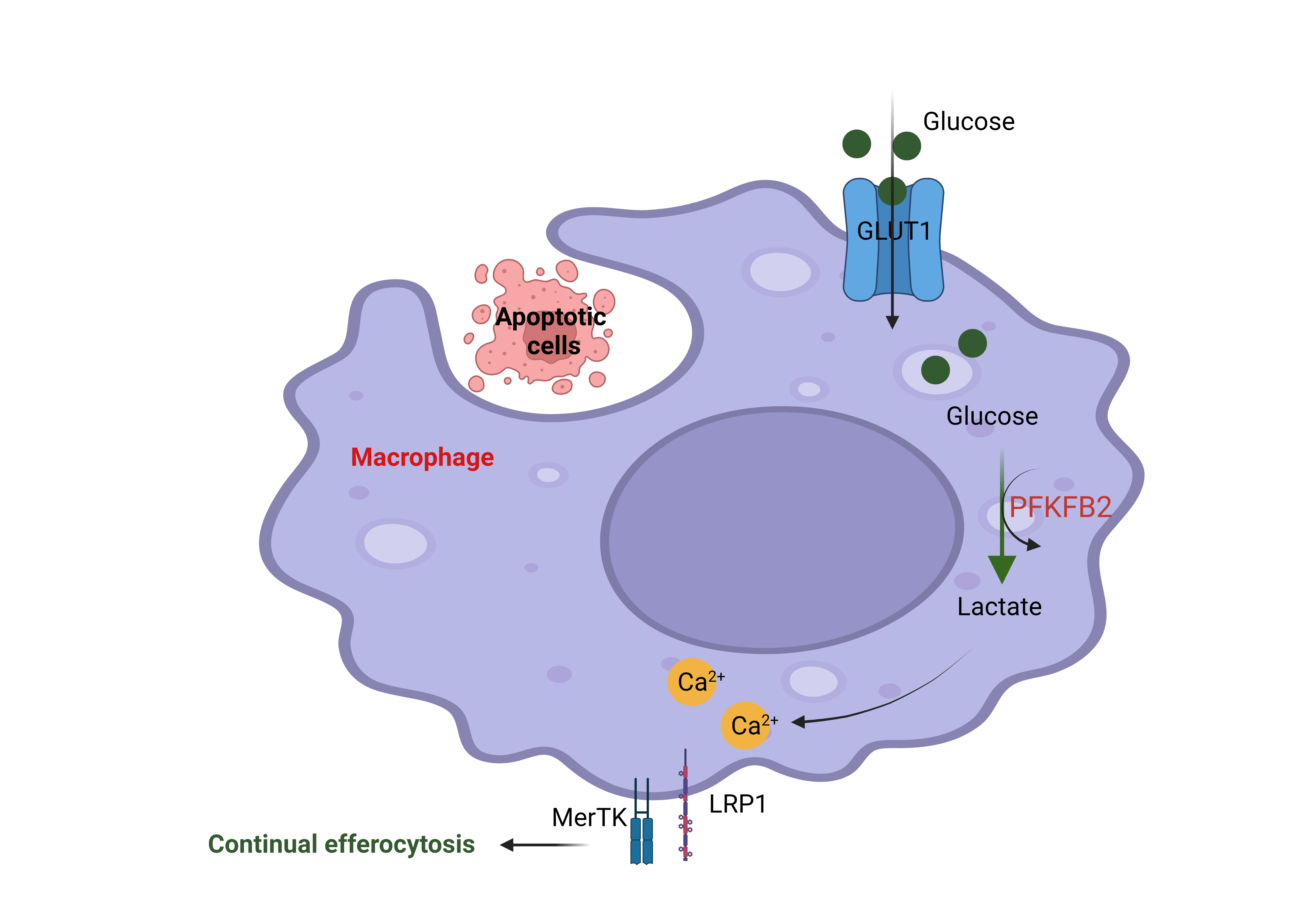
Figure 2: The engulfment of apoptotic cells by macrophages through efferocytosis increases glucose uptake via the membrane transporter GLUT1. Glucose is broken down into lactate through glycolysis, and this process is boosted by efferocytosis through activation of the enzyme 6-phosphofructo-2-kinase/fructose-2,6-bisphosphatase 2 (PFKFB2). Lactate subsequently increases cell surface expression of the efferocytosis receptors MerTK and LRP1. These efferocytosis receptors facilitate the  subsequent uptake and degradation of other apoptotic cells in the tissue. This figure was created using Biorender.

MerTK and LRP1 are so-called “efferocytosis receptors” that allow the macrophages to  bind to dead cells before they can engulf and degrade them. The current study found that the production of lactate leads to an increase in MerTK and LRP1 on the cell surface in a calcium-dependent manner to drive continual removal of dead cells (Figure 2). The authors mentioned that lactate promotes an efferocytosis-induced calcium-raising mechanism that could be involved in the mitochondria division. The mechanism of how lactate promotes the increasing of calcium is not well understood and needs to be explored more in the future. This finding provides potentially new therapeutic strategies for improving cell death clearance such as targeting an endogenous inhibitor of PFKFP2. This novel finding was published in Nature Metabolism in February 2023. 

Reviewed by: Maaike Schilperoort , Erin Cullen, and Sam Rossano

Using AI to identify high-risk patients in home health care

The population is aging. Over the past two decades, life expectancy in the United States has increased by more than five years to approximately 80 years in 2020 and is projected to further increase to over 85 years in 2060. The progressive rise in life expectancy leads to a growing share of older people in society, which increases the demand for health care services including home health care (HHC). HHC is defined by medical services that are provided in a patient’s home, usually by skilled nurses. An important aspect of HHC is the identification of patients who are at high risk for emergency care. A substantial fraction of HHC patients has to visit the emergency room or is admitted to the hospital during the HHC period. Strikingly, up to 30% of these events could be avoided by accurate risk prediction and preventative care. For example, patients identified as high-risk can be monitored more closely and treated with additional medications to prevent adverse health outcomes.

The research of Columbia postdoc Jiyoun Song is aimed at improving risk prediction in the setting of HHC. In recent work published in the Journal of Advanced Nursing, Dr. Song and colleagues performed cluster analysis, an unsupervised machine learning method to aggregate available patient data into groups. Such a clustering method is useful in identifying patterns of risk factors that interact with each other, rather than examining individual risk factors. The analysis was performed on structured data from electronic health records, that include patient characteristics such as socio-demographics and health conditions, as well as unstructured data from clinical notes written by nurses. The approach of Dr. Song and colleagues to use data derived from these clinical notes is quite innovative. They extracted risk factors from these notes through the artificial intelligence (AI)-based tool of natural language processing. Natural language processing can be used to extract meaning from text written by a human, which forms the foundation of AI chatbot systems such as the recently developed ChatGTP. The use of clinical notes as an additional source of data significantly improves risk prediction in the HHC setting, as previously shown by Dr. Song.

Through AI-assisted cluster analysis of both the structured and unstructured data, three clusters of risk factors were identified with distinct characteristics: (1) impaired physical comfort with pain, (2) high comorbidity burden (i.e., a person is suffering from more than one physical disease or condition at the same time), and (3) impaired cognitive/psychological and skin integrity. Classifying patients in these three categories could help tailor individualized preventive strategies. For example, a pain management strategy may work best for patients in cluster 1, while patients in cluster 3 may benefit mostly from psychological counseling and wound management. The study also found that these clusters are associated with the frequency and timing of emergency room visits. Patients that fall into cluster 3 have the highest need for emergency care, with 15.7% of patients being hospitalized or visiting the emergency department within the first 60 days of HHC (see Figure below).

AI-assisted cluster analysis of both structured data (i.e., electronic health records) and unstructured data (i.e., clinical notes) identified three clusters of risk factors with distinct characteristics. The risk of hospitalizations or emergency department (ED) visits is different for each cluster. Such a cluster-based analysis is useful for identifying high-risk patients in home health care and implementing preventative strategies. © 2022, Maaike Schilperoort

The results from this study suggest that implementation of cluster-based risk prediction models into early warning systems could reduce the likelihood of HHC patients being admitted to the hospital. This illustrates the potential of AI-based methods for clinical risk prediction. The use of AI is getting increasingly popular and is now also implemented in various other aspects of healthcare, such as in-hospital decision making, predicting treatment benefit, and personalizing medicine. To what extent will AI be incorporated in our healthcare system? Only time will tell.

Reviewed by: Jiyoun Song, Pei-Yin Shih, Trang Nguyen, and Sam Rossano

Seeing more of the Unseen – using vibrational contrast and MARS to improve microscopy

Biomedical imaging is an important tool in science because it allows scientists to see what may not be visible to the human eye. Using light within the visible spectrum, microscopy allows us to see cells and their functional subunits called organelles, which can be thought of as the internal organs of a cell. We can also visualize certain proteins that may be expressed within certain organelles using fluorescence microscopy. With fluorescence microscopy, proteins in tissue or cells are tagged with light emitting markers, called fluorophores. Fluorophores make proteins under the microscope light up like fireflies on a dark summer night. Different color fluorophores can be used simultaneously to image different proteins at once, however this is limited by the number of colors available in the visible light spectrum. This means that with fluorescence imaging on a confocal microscope, there are a limited number of proteins that can be imaged within a given sample. 

That’s where MARS comes in (not the planet!)! Manhattan Raman Scattering (MARS) is a special dye pallet that, combined with signals from an electronic pre-resonance Stimulated Raman Scattering microscopy (epr-SRS), creates a very sensitive way to probe, visualize and image organelles with vibrational contrast, as opposed to just light contrast. Vibrational contrast detects molecules based on their chemical properties. For example, if a probe molecule has a double bond, it will have a different vibrational frequency than a molecule with a single bond. With this technology, you can differentiate cellular targets by using dyes/probes that vary by light and vibrational signals, making these techniques very sensitive. However, these MARS dyes are difficult to chemically synthesize, and there were initially only a limited number of usable MARS dyes.

Columbia postdoc Dr. Yupeng Miao and colleagues published an article in 2021, summarizing their development of new MARS dyes that have different properties that are easier to synthesize and can visualize even more of the cell’s proteins under the microscope at once! The research contributed 30 new MARS probes that can specifically label various proteins of interest within a given sample.

Before synthesizing these new MARS probes, the researchers designed and simulated models for each potential dye. For the design, they used a similar foundation to the previous MARS probes, but included some adjustments like changing the core atom or substituting stable isotopes throughout the molecule. The results from the design models gave the researchers confidence that they could synthesize these edited molecules to expand the list of available MARS probes.

Indeed, they expanded the list of probes by developing 30 new molecules that are able to label specific cell organelles and functions. For example, MARS probes were used to image subcellular structures including the protein alpha-tubulin, which is a protein within microtubules that provide structural support to the cell, as well as fibrillarin, which is a protein that is used as a nucleoli marker. MARS probes were also shown to successfully target the cell membrane, mitochondria, lysosomes, and other lipid structures within the cell. Even more exciting – this technology allows researchers to probe each of these cellular structures simultaneously, moreso than can be done with current fluorescent microscopy methods. This means that the new MARS probes can be used to image multiple cellular markers within the same sample!

With this technology, scientists can now see even more of the unseen, which can expand our knowledge on cellular (dys)function in health and disease.

Edited by: Maaike Schilperoort, Trang Nguyen

The Importance of Consistent Sleep for Memory Retrieval at the Neural Level

Sleep helps us remember the details of past events more clearly. When we sleep, neural mechanisms facilitate the consolidation of memories formed during the waking day. Specifically, memories are temporarily stored in a brain structure called the hippocampus. During the consolidation process, memories are replayed and integrated into long-term storage centers in the neocortex of the brain. Poor sleep impairs sleep-based memory consolidation and memory retrieval. In other words, when our sleep is fragmented, our memory is less clear. 

One way to assess the clarity of a memory is to measure neural similarity, or the overlap between patterns of neural activity.  My colleagues and I presented participants with a series of word pairs to remember while we recorded their neural activity using electroencephalography. We used this task to measure neural activity when participants studied (i.e., encoded) and were tested on (i.e., retrieved) the word pairs. The overlap between their neural patterns for a given word pair at study and test is an index of neural similarity.

Interestingly, we found that sleep quality was associated with neural activity for word pairs that were paired differently. When people had more consistent sleep quality from night-to-night (measured with wrist-worn monitors), they had greater neural similarity when they correctly rejected word pairs that were paired differently. For example, if they saw the pair “wing – clock” during the study period and correctly identified “fork – clock” as a different pairing at test, they demonstrated higher neural similarity. 

There were several strengths of the study. We used an objective measure of sleep quality — wrist-worn monitors. We also measured sleep quality for seven nights, which allows for assessing night-to-night sleep variations. Our participants were racially and ethnically diverse people across the adult lifespan. However, our study was limited by its small, convenience-based sample of participants (74 people) and cross-sectional design. We cannot determine if poorer sleep causes lower neural similarity with this data. 

Taken together, our study suggests that memory integrity, or the ability to clearly remember the details of past events, may be linked with consistent sleep patterns. Thus, in addition to sleeping for enough time, sleep consistency also contributes to better memory retrieval. 

Edited by: Trang Nguyen, Pei-Yin Shih

Engineered bacteria enhanced the current therapeutics in lung cancer

Lung cancer is the second most common type of cancer and is responsible for the most cancer-related death in the U.S. The American Cancer Society reports that more than 235,000 people were diagnosed with lung cancer in 2021. There are three major types of lung cancer: non-small cell lung cancer (85% of cases), small cell lung cancer (10% of cases), and lung carcinoid tumor (5% of cases). The causes of lung cancer include but are not limited to smoking, secondhand smoke, exposure to certain toxins, and family history. The symptoms include cough with blood, chest pain, wheezing, and weight loss when the cancer is in the advanced stage. Depending on the type of lung cancer and what stage it has progressed to, the treatment will be different. Broadly the treatment involves surgical resection, radiation, chemotherapy, targeted therapy and immunotherapy. However, to treat this complex disease researchers are always looking for new and improved treatment modalities.

A research group at Columbia Engineering led by Dr. Dhruba Deb in the lab of professor Tal Danino developed a new therapeutic to treat non-small cell lung cancer (NSCLC) by combining an engineered bacteria with targeted therapy to enhance the treatment efficacy without any additional toxicity in laboratory animal models. This finding was published in Scientific Report on December 13, 2022.

By engineering a toxin named theta (θ) toxin in the bacteria S.typhimurium and by testing the response of a variety of NSCLC cells to this engineered bacteria, the research group found that θ toxin can kill a variety of NSCLC cells even with different genetic background such as mutated growth factor receptor like KRAS or EGFR, the most common mutations found in NSCLC. The research group also administered locally live S.typhimurium expressing theta toxin (Stθ) in NSCLC tumor cells in the mouse model and found a 2.5-fold reduction of tumor growth within a week compared to the control group. 

With the success of testing live S.typhimurium expressing theta toxin (Stθ) in mouse model and no toxicity  found in the peripheral organs, the research group tested whether using the engineered bacteria could enhance the efficacy of the standard of care chemotherapies as well as small molecular inhibitors. To identify potential drugs to combine with Stθ, the authors used RNA-sequencing. This helped to pinpoint which biochemical pathways in NSCLC cells were helping the cells to survive the Stθ treatment. To overcome this ability of NSCLC cells, the researchers blocked those biochemical pathways with drugs and eliminated the NSCLC cells. For example, one of the drugs, MK2206 when paired with Stθ treatment, blocks the NSCLC cells’ ability to survive via biochemical signaling of phosphorylated AKT that promotes survival and growth in response to extracellular signals. Key proteins involved are PI3K (phosphatidylinositol 3-kinase) and Akt (protein kinase B)..  The research group also tested the combination treatment of MK2206 and Stθ in a mouse model. They found that the combination treatment of Stθ bacteria and MK2206 suppressed the tumor growth efficiently compared to treatment with only Stθ or only MK2206. Moreover, with lower dose of bacteria and drug use, they could observe similar treatment results and could possibly avoid  the activation of the immune system caused by high dose of bacteria treatment. Taken together, this combination treatment is a potential therapeutic for the NSCLC. 

There are several limitations in this study that need to be addressed before entering the clinical trial. First, the authors used a small number of animals per cohort in the in vivo study, so they plan to expand their study to assess the overall survival upon treatment. Second, the toxins themselves are not selectively targeted to the cancer cells, so they need to develop a selective delivery method to avoid the systemic toxicity. In the laboratory animal models, the local administration of the live bacteria acted as a selective delivery. But, further studies are necessary to use this live bacteria in human clinical trials. Overall, this study opens up new treatment options for patients diagnosed with the NSCLC.

Reviewed by: Sam Rossano, Margarita T. Angelova

A continuous war between DNA elements shapes genome evolution

While genomes include the totality of genes that determine an organism’s biological identity, genes can only be a tiny fraction of the genome. In humans, and in many other species, DNA contains multiple diverse regions. One example are transposons, or mobile genetic elements, which are parts of DNA that can move in new places in the genome (Figure 1). The “genomic walks” of transposons are potentially harmful, since when they “jump” within a gene, they trigger mutation and loss of that gene. Cells are constantly evolving new strategies to keep transposons under control. In response, transposons adapt and arm themselves with new strategies to escape cellular control.

Sometimes, a mobilized transposon hijacks additional DNA information that it transfers to the new location with itself. When this additional DNA happens to be a gene providing survival advantage, not only the recipient increases its gene pool and survival capacities but the transposon also increases its chances to propagate. This process of transmission of information between different species is termed horizontal gene transfer and represents a major driver of genome evolution across all domains of life. Transposons have been key players in this process.

Figure 1. Schematic representation of a DNA transposon and its movement between two DNA molecules. Image created with BioRender.com.

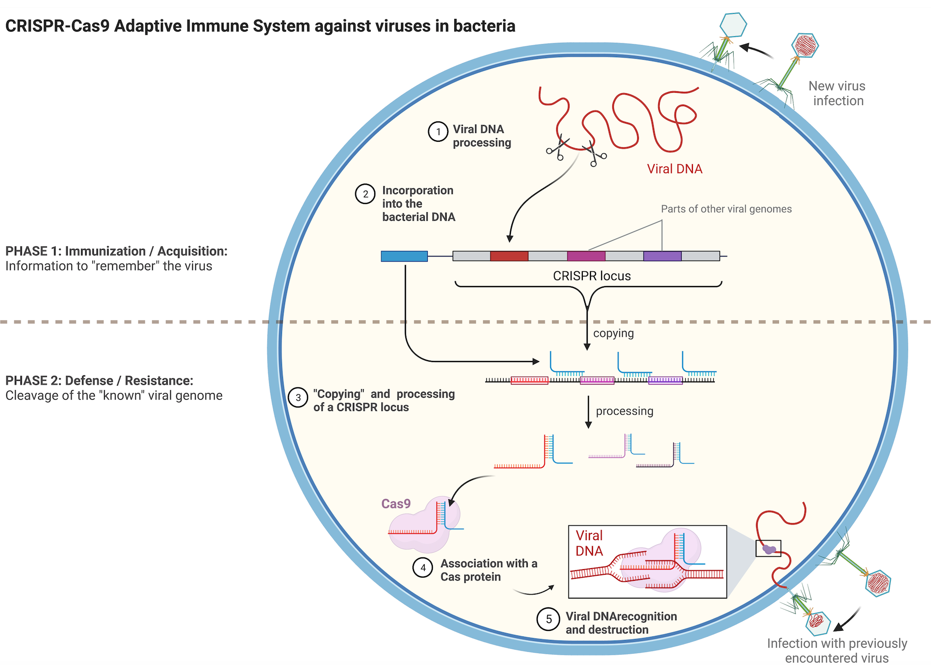
Transposons can be considered as reminiscent of ancient viral infections that managed to integrate in a host cell genome but later lost the capacity to completely exit the cell. Bacteria have evolved a fascinating mechanism to remember viruses that they have already encountered. This acquired immunity involves a family of DNA sequences named CRISPR and their associated protein partners Cas. The CRISPR sequences are fragments of DNA derived from different viruses that have infected the bacteria and were integrated in the bacterial genome, creating a footprint of previous infections (Figure 2, upper part). When cells are infected, they use the CRISPR catalog and compare it to the invading DNA, helping it to recognize recurrent viruses and destroy them more rapidly. Cas proteins participate in the degradation of the viral DNA. (Figure 2, lower part).

Advances in research showed that the CRISPR-Cas complexes can be modified to edit genes in different organisms. To do this, part of the complex is changed to lead the Cas protein to a gene of interest, instead towards a viral genome. This system of gene editing has already found numerous important applications ranging from basic biology research to disease treatments and development of new technologies. The discovery of CRISPR-Cas was acknowledged with the 2020 Nobel Prize in Chemistry to Emmanuelle Charpentier and Jennifer Doudna.

Figure 2. Schematic representation of the CRISPR-Cas9 adaptive immune system of bacteria. Briefly, upon infection the viral DNA is fragmented and part of it is integrated in a special region of the bacterial genome, the CRISPR locus. CRISPR sequences get copied into shorter RNA molecules that carry parts of sequences identical to the sequence of a certain virus. Once copied, these short RNAs form complexes with Cas proteins and serve as “guides” for them towards potential complementary viral DNAs. The viral DNA is destroyed by the Cas protein if it can hybridize with the short RNA from the CRISPR locus. Image created with BioRender.com. 

Interestingly, researchers have found an intriguing interconnection between transposons and the  CRISPR–Cas defense system. In a striking example a bacterial transposon has hijacked some of the genes of a CRISPR-Cas system and uses those genes for its own propagation in genomes. These transposons are called CRISPR-associated transposons. Such widespread exchange of genes is caused by the never-ending arms race between the transposons and their hosts’ defense systems. In the recent publication of Hoffman and colleagues, five Columbia postdocs Minjoo Kim, Leslie Y. Beh, Jing Wang, Diego R. Gelsinger, and Jerrin Thomas George collaborated and brought significant insight in the functioning of one CRISPR-associated transposon.

In their publication, the authors monitored in detail the formation of  this RNA-guided transposon and its associated complexes, which enabled them to resolve distinct protein recruitment events that take place before the integration of the transposon. They also found that even if initially hundreds of non-desired genomic sites are targeted for integration at the end only few of those sites recruit the whole transposon machinery that is required for integration at this genomic location. This discovery offered insights into how the potential target sites in the host genome are identified, screened and approved for integration, allowing the transposon system to be specific.

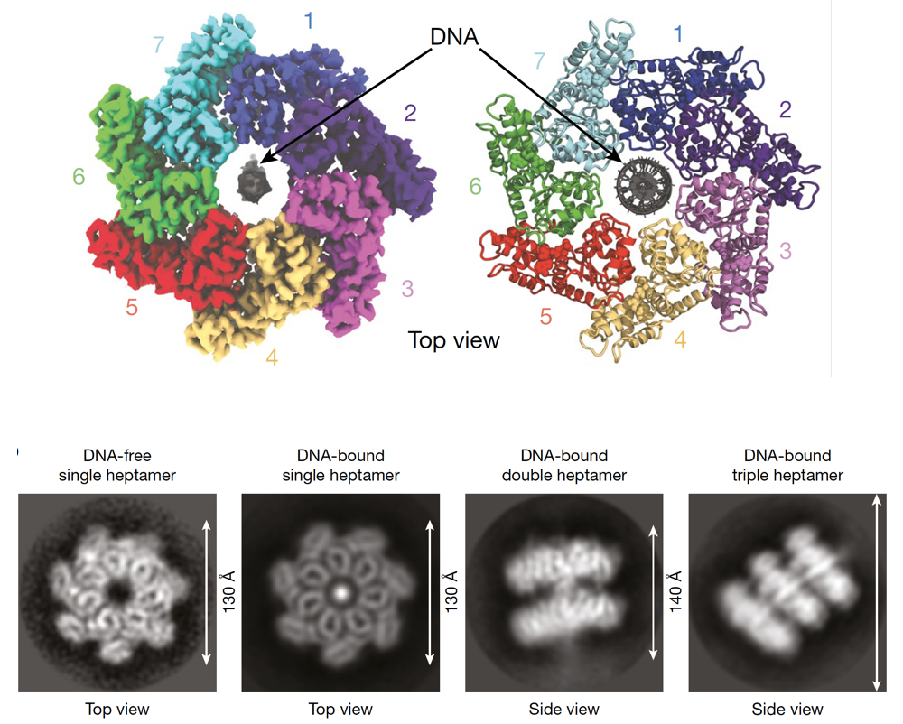
To advance the understanding of interactions responsible for the assembly of the transposon associated proteins, the authors determined the structure of one of the interacting proteins, named TnsC and found that it is forming rings of seven molecules of TnsC that can pass DNA through the central pore of the ring. This helps to correctly position DNA for the following integration (Figure 3). The resolved molecular structure also allowed to gain clarity on how TnsC mediates the communication between the proteins in this transposon complex that are responsible on the one hand for the targeting and on the other hand for the integration at a specific genomic location. Their results pointed to TnsC as the proofreading checkpoint that ensures the specific selection of genomic sites for transposition.

Figure 3. Upper part: Model of the 7 molecules of TnsC forming a ring through which DNA is passed. Lower part: Representative experimental result from Hoffmann et al. showing different configurations and views of the DNA-TnsC complex. Figure adapted from the original publication. 

In summary, the paper not only deciphers the molecular specificity of consecutive factor binding to genomic target sites in this interesting process of RNA-guided transposition, but the resolved detailed structure also provides valuable information for the development of future biotechnologies in the field of programmable and specific integration of DNA in desired genomic locations. Such technology differs from the above-described original CRISPR-Cas system currently used for genome editing, because it has the potential to be less mutagenic, as well as because it provides the opportunity to insert much longer pieces of DNA in a desired location. RNA-guided transposases hold tremendous potential for future biotechnological and human therapeutic applications and will without a doubt accelerate novel discoveries. Find out more in the original publication.

Reviewed by: Trang Nguyen & Maaike Schilperoort

A high-tech device to effectively deliver drugs to tumors in the brain

Brain and other nervous system cancer is the 10th leading cause of death for men and women. Around 18,280 adults died from primary brain and central nervous system tumors in the United States in 2020. Glioblastoma is the most common malignant brain and other CNS tumors and median survival is only 12-15 months.

Why is brain tumor hard to treat? It is due to the blood brain barrier (BBB), a specialized network of blood vessels and cells that shields and protects the central nervous system against circulating toxins or pathogens that could cause brain infections. However, the impenetrability of the BBB also makes it difficult to treat tumors in the brain compared  to those in other organs. Patients with brain tumors have to receive higher doses of chemotherapy to penetrate the BBB and ensure an adequate amount of medication reaches into the brain and kills the tumor cells. The higher dose of chemotherapy will lead to the toxicity to the normal cells, which can result in serious side effects and even death of the patient. To overcome the BBB, scientists have tried to develop many different methods to deliver drugs effectively to the brain so that lower doses of chemotherapy can be used.

Over the last decade, Drs. Bruce and Canoll’s laboratory at the Columbia University Medical Center has been developing a new method to directly administer drugs to the site of the brain tumor, which they call convection enhanced delivery (CED). In CED, a small pump is implanted into the abdomen and connected to a thin catheter under the skin. Wireless technology is used to turn the pump on and off and control the flow rate of medicine that seeps in the tumor tissue.

In a recent study with the CED device, Dr. Bruce used topotecan, a drug that is toxic to glioblastoma cells, to treat five patients who were at least 18 years old with recurrent brain tumors. The patients were infused with topotecan for 48 hours, followed by a 5–7 day washout period before the next infusion, with four total infusions. Patients went about their normal routines at home while treatment continued without any severe side effects. After the fourth infusion, the pump was removed and the tumor was resected. This method is in the early phase of clinical trials (phase 1b) and will be expanded to a larger scale of patients due to test the safety and efficacy of the therapy for recurrent glioblastoma. This novel chemotherapy delivery strategy overcomes the limitation of drug delivery in patients with glioma. The results from this study have recently been published in Lancet Oncology 

There are two limitations in this study. First, there is no comparison group for determination of definitive survival benefit. Second, there is no way to assess the disease progression and treatment response due to effects of local drug infusion and surgical resection. However, in the locally delivered therapy (CED method), the authors used patients as their own control by performing pre-therapy and post-therapy MRIs and PET scans. The CED device effectively gets through the BBB to kill the brain tumor so new classes of drugs and targeted compounds could potentially be used such as high-molecular-weight compounds or viruses.

Reviewed by: Pei-Yin Shih, Maaike Schilperoort

Follow this blog

Get every new post delivered right to your inbox.