“super-SOX” hits iPSC research out of the park

The human body contains trillions of cells, which have extremely diverse functions. Remarkably, every one of these cells has the same genetic code. This diversity is possible because of the developmental process of differentiation. As an organism grows, cells take on traits that are useful in a particular context. For example, skin cells produce a protein called keratin that makes skin tough in order to protect the rest of the body from the outside world. Other cells don’t produce keratin because it would hinder their function. Once cells have differentiated and taken on a particular role, they can only generate more of the same kind of cell – skin cells only divide into more skin cells. This is because sections of DNA are hidden away during differentiation and stay hidden after the cell divides, ensuring that all the cells in a particular organ (like the skin) act like they’re supposed to.

To turn one type of differentiated cell into another was once thought to be impossible. The only cells that can generate any type of cell in the body are pluripotent stem cells (PSCs). PSCs have many potential applications in science, medicine, and industry, including disease modeling and drug testing as well as cell-based therapies and biotechnology. The opportunity to study PSCs can also enable scientists to answer basic questions about mammalian development. 

In 2006, Japanese researchers Takahashi and Yamanaka discovered that PSCs actually can be created from differentiated cells. Doing so requires just four key proteins: Oct4, Sox-2, Klf4, and cMyc. These proteins together are referred to as the Yamanaka factors. The discovery that the Yamanaka factors could turn a differentiated cell into a PSC was incredibly exciting to the scientific community, earning Dr. Shinya Yamanaka and a colleague the Nobel Prize in Physiology or Medicine in 2012.

Although this discovery created a broad array of new possibilities in science, it turns out that making PSCs in a laboratory is tough. Making healthy, high-quality PSCs from different species is even tougher. Induced pluripotent stem cells (iPSCs) have only been generated from a few species, and some cell lines create healthier iPSCs than others. A clearer understanding of how the Yamanaka factors interact with DNA and each other could reveal ways to make iPSC generation more robust and consistent, and realize the exciting potential of iPSCs. 

This challenge was taken on by a research group led by Drs. Sergiy Velychko and Caitlin MacCarthy in collaboration with CUIMC postdoc Dr. Vikas Malik and other teammates around the world. The group published a study this month in Cell Stem Cell describing a new protein based on the Yamanaka factor Sox2. They aptly call this protein “super-SOX” based on a number of studies demonstrating its ability to better generate iPSCs. The experiments also uncovered basic principles of pluripotency and development.

While three of the Yamanaka factors can be replaced with other proteins from the same family to generate iPSCs, Oct4 is the only member of its family (the POU family) that can induce pluripotency. The researchers set out to understand this oddity, with the goal of revealing the molecular interactions that lead to pluripotency. They first found that while a different member of the POU family could not generate iPSCs with the Yamanaka factor Sox2, it could generate them with a mutant protein based on another member of the SOX family, Sox17. 

Next, the researchers created a library to test different Sox2-Sox17 chimeras – mutant proteins that include components from both Sox2 and Sox17. Amino acids are the most basic building blocks of proteins, and surprisingly, the researchers found that a chimera with only one different amino acid from Sox2, Sox2AV, stabilizes the interaction between Sox2 and Oct4 and increases DNA binding efficiency. Oct4 is distinguishable from other members of the POU family by a negative charge in its “linker” domain. This negative charge allows Oct4 to form “salt bridges” connecting it to Sox2. Swapping the Sox2 amino acid alanine for the Sox17 amino acid valine promotes the formation of these bridges, encouraging Sox2AV and Oct4 to interact and bind to DNA together. The stronger connection between these two proteins dramatically improved pluripotency. 

Photo of healthy adult mice generated entirely from iPSCs and schematic of how outcomes change dramatically after replacing Sox2 with Sox2AV (Adapted from McCarthy et al., 2024).

The researchers hypothesized that the improvements in DNA binding they observed could promote the development of iPSCs into mature organisms. Live mice and rats have previously been generated from iPSCs, but these animals rarely survive to adulthood. Replacing Sox2 with Sox2AV – a simple change of one amino acid – when generating iPSCs dramatically increased mouse survival. 10/10 different cell lines treated with Oct4, Sox2AV, Klf4, and cMyc were able to produce live all-iPSC mouse pups, which only 3/8 cell lines treated with all the traditional Yamanaka factors were able to achieve. In the best-performing Sox2AV line, 43.3% of all-IPSC mice became healthy adults, compared to only 15.2% from the best-performing Sox2 line.

A further study found that a more complex chimera of Sox2 and Sox17 (Sox2-17) could improve iPSC generation in five different species: mice, cows, pigs, monkeys, and humans. These species were chosen for their potential to bring iPSC technologies to either biomedical research or industry – for example, iPSCs from livestock species could be key to producing lab-grown meat. Non-human primates and livestock species are less established in iPSC research, but the invention of Sox2-17, or “super-SOX”, opens many doors for future uses of iPSCs from these species. 

One barrier to creating iPSCs from human cells is age – differentiated cells from adults are highly resistant to becoming iPSCs. Strikingly, the group found that although the traditional Yamanaka factors could not produce iPSCs from skin cells of certain Parkinson’s disease patients, replacing SOX2 with the chimeric SOX2-17 could produce iPSCs from these samples.  Parkinson’s disease is characterized by the selective death of neurons in a brain region important for motor coordination called the substantia nigra, and could theoretically be treated by replacing lost neurons with iPSCs differentiated into neurons with a particular patient’s genetic code. The finding that SOX2-17 improves iPSC generation in cells from Parkinson’s disease patients demonstrates an exciting potential medical application for this discovery. 

The paper goes on to provide an explanation for these exciting findings. Prior to this study, it was known that although Oct4 is necessary for inducing pluripotency, too much Oct4 impairs iPSC generation. Free Oct4 encourages proliferation, which is actually detrimental to inducing pluripotency. Here, the researchers found that the stronger bond between Oct4 and Sox2AV or Sox2-17 lowers the proliferation rate, which improves the health of the cells that are generated. They went on to show that the bond between these two proteins is a main driver of the process of reverting a differentiated cell to a “naive” iPSC, which broadens the possibilities for the cell’s future development. 

This study makes a significant practical step toward realizing the potential of iPSCs. Simultaneously, it answers longstanding questions regarding how exactly the Yamanaka factors induce pluripotency, showing that the interaction between Oct4 and Sox2 has a central role. These findings have exciting implications for iPSC research, a field at the very forefront of science and medicine. 

Reviewed by: Trang Nguyen, Giulia Mezzadri, Carlos Diaz, Vikas Malik


Make new friends or keep the old? A novel brain circuit explains social novelty preference in mice

Researchers have discovered a pathway in the brain that may dictate whether we decide to interact with a friend versus a stranger. “Corticotropin-releasing hormone signaling from prefrontal cortex to lateral septum suppresses interaction with familiar mice”, was published this month in the highly regarded journal Cell. The paper emerged from a collaboration between scientists at the Zuckerman Mind Brain & Behavior Institute at Columbia University and colleagues at the University of Washington in Seattle, Washington and the Instituto de Neurociencias CSIC-UMH in Alicante, Spain. Columbia postdoc Ramon Nogueira contributed to the work. 

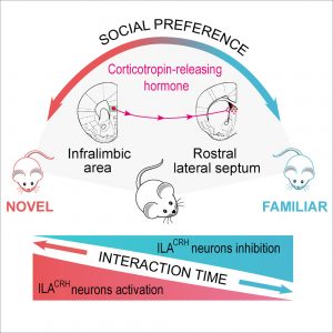
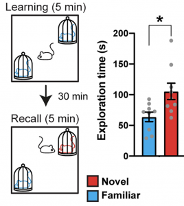
A diagram demonstrating Social Novelty Preference in mice. Two mice are in enclosures on opposite sides of an arena. A third mouse, free to explore the arena, chooses to spend more time with a mouse it does not know than a mouse it has met before.
Fig. 1 – Diagram of the task to assess Social Novelty Preference in mice. A mouse with typical social memory and an intact connection between CRH-releasing neurons in the ILA and CRH-receiving neurons in the rLS will spend more time interacting with the novel mouse than the familiar mouse. (Adapted from de León Reyes et al., 2023, Figure 3)

Social novelty preference in mice was first described in the scientific literature 20 years ago, and it is regularly used to assess social memory for research purposes. A typical mouse, when presented with a mouse it knows and a stranger, will spend more time with the familiar mouse (Fig. 1). Until this paper was published, it was unclear what brain regions or pathways influence whether mice choose to interact with novel or familiar mice. Scientists theorized that the lateral septum, a relay center that connects regions of the brain associated with thinking and memory, and the prefrontal cortex, which controls decision-making, might play a part in the social novelty preference. Both regions are also known to be involved in social behavior, and evidence suggested that there may be a neural pathway leading from the infralimbic area (ILA) in the prefrontal cortex to the rostral lateral septum (rLS), a specific region within the lateral septum. The researchers set out to determine how these two regions interact with each other and whether this interaction plays a role in social novelty preference. 

Using a comprehensive and elegant set of experiments to manipulate neuron function in the two regions, the researchers discovered that a population of inhibitory neurons in the ILA connects to the rLS, and that this pathway is crucial for the mice to exhibit the typical social novelty preference. The ILA neurons contain the neurotransmitter corticotropin-releasing hormone (CRH), which is best known for its role in stress and anxiety. Neurons in the rLS have receptors for CRH, but it was previously not known that CRH-containing neurons in the ILA communicate with CRH-receiving neurons in the rLS. When the researchers inactivated the CRH-containing neurons in the ILA, they found that mice failed to exhibit the typical social novelty preference. Next, they asked whether the CRH-receiving neurons in the rLS were also necessary for social novelty preference. They found that removing the CRH receptors from these neurons erased the social novelty preference, just like inactivating the CRH-releasing ILA neurons. Finally, they recorded the activity of CRH-releasing ILA neurons and saw that these neurons were more active when interacting with a familiar mouse than a novel mouse, indicating that these neurons act on the rLS to discourage interactions with familiar mice. 

Graphic showing a pathway through which corticotropin-releasing hormone is sent from the infralimbic area to the rostral lateral septum of the mouse brain. When this pathway is active, mice spend more time with novel rather than familiar mice. When this pathway is inactive, mice spend more time with familiar rather than novel mice.
Fig. 2 – Graphical Abstract of “Corticotropin-releasing hormone signaling from prefrontal cortex to lateral septum suppresses interaction with familiar mice” by Noelia Sofia de León Reyes et al., 2023.

To further probe the relevance of this pathway to social dynamics, the researchers assessed whether CRH may be responsible for the social novelty preference appearing in young mice. While adult mice prefer to interact with novel mice, mouse pups less than two weeks old prefer to interact with their mothers and littermates rather than unknown adult females or pups from other litters. During this same developmental time period, the population of CRH-releasing neurons that connect to the rLS steadily grows. When CRH was depleted from these neurons, the young mice failed to exhibit the typical shift from a preference for social familiarity to a preference for social novelty. This may be an evolutionary mechanism that encourages young mice to stay in safe, familiar environments, then emboldens adult mice to explore novel environments, collect food, and procreate. 

While the social novelty preference in mice does not have a direct analogue in humans, this research may provide clues for understanding and treating social anxiety disorders and social phobias. The human prefrontal cortex and septal regions also respond to social information, and CRH is known to be involved in anxiety disorders including social phobias. The researchers suggest that low CRH levels in the prefrontal cortex may prevent people from seeking social novelty, and that this mechanism could be responsible for social anxiety disorders. The field of neuroscience is just beginning to understand the biological underpinnings of social interaction, and new methods give scientists ever expanding ability to learn about this exciting field. 

Reviewed by: Trang Nguyen and Martina Proietti Onori

A new advance in understanding the development of binocular vision

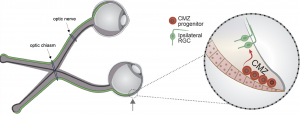
Have you ever wondered how two eyes can work together to create a single image? This occurs through a complicated mechanism called binocular vision, or stereopsis. When light hits the eye, photosensitive cells called rods and cones translate that light into an electrical signal, which they communicate to retinal ganglion cells (RGCs). RGCs have long axons that transmit visual signals to neurons in the brain. These axons are initially carried from the eye to the brain in a bundle called the optic nerve. Each eye has an optic nerve, and the two optic nerves meet at an intersection called the optic chiasm. At the optic chiasm, some of the RGC axons from each optic nerve cross to the opposite, or contralateral, side of the brain and the rest stay on the same, or ipsilateral, side. This process allows the part of the world that the left eye can see and the part that the right eye can see to overlap in the center, forming one cohesive image. 

Figure 1. The role of the CMZ in ipsilateral RGC generation. On the left, a diagram shows axons either remaining on the ipsilateral side or crossing to the contralateral side of the brain at the optic chiasm, a process which makes binocular vision possible. Ipsilaterally projecting RGC axons are shown in green, and contralaterally projecting RGC axons are shown in black. On the right, CMZ progenitors are shown differentiating into ipsilaterally projecting RGCs (modified from Slavi et al., 2023).

When binocular vision does not develop normally, depth perception can be impaired. This is often the case in albinism, a genetic condition characterized by low levels of melanin that occurs in humans and other mammals. While melanin is most widely known for its contributions to skin color in humans, it also plays an important role in the development of the visual system. The exact nature of this role is still being uncovered. Recent work emerging from a collaboration between the Mason-Dodd lab and the John lab at Columbia University’s Zuckerman Institute, led by Postdoctoral Research Scientist Dr. Nefeli Slavi and Associate Research Scientist Dr. Revathi Balasubramanian, used albino mice to identify why deficits in depth perception might occur in albinism. The study, published in the leading neuroscience journal Neuron, also advanced our understanding of the development of binocular vision generally. 

Most RGCs are formed in a location called the neural retina during development, but recent research has uncovered a prominent role for another source of RGCs called the ciliary margin zone (CMZ). The CMZ produces mostly ipsilaterally projecting RGCs, which are present at abnormally low levels in albinism. Based on this existing research, Drs. Slavi and Balasubramanian and their co-authors decided to investigate why these cells are not produced at typical levels in albino mice. First, the group found that there were fewer cells in the actively dividing phases of the cell cycle in the CMZ in albino mice. Further investigation revealed that this deficiency in cell division led to a lower number of ipsilaterally projecting RGCs in these mice. They traced these deficits to decreased expression of a protein called CyclinD2, which is known to encourage cell division. To further investigate the role of CyclinD2 in cell division in the CMZ, the researchers used mice that were pigmented (not albino) but did not express CyclinD2 in the CMZ. These CyclinD2-deficient mice had the same impairments in cell division as the albino mice.

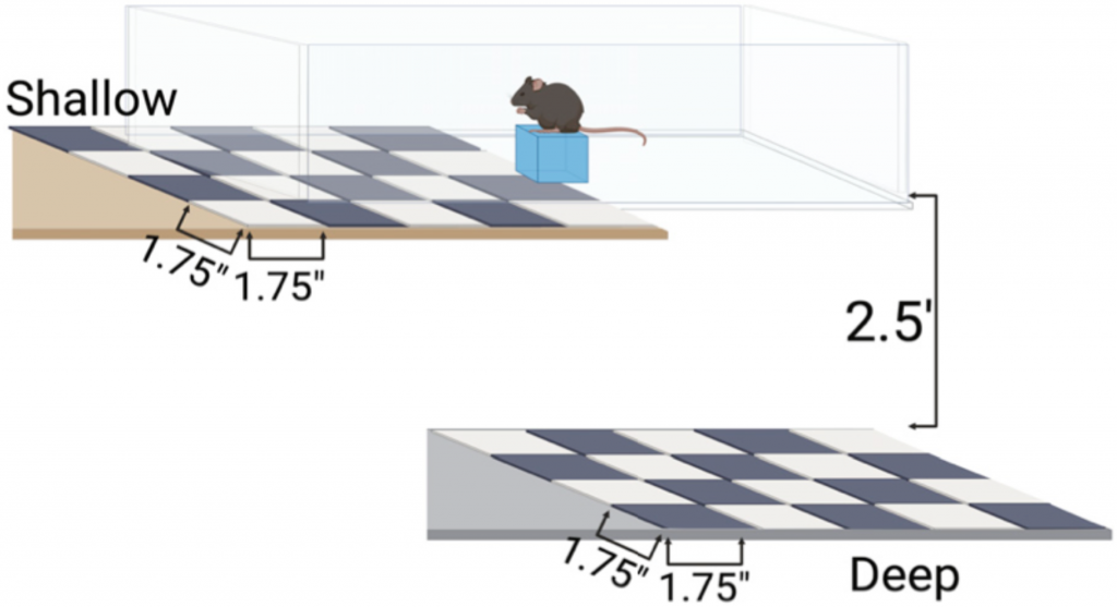
Next, the researchers tested the impact of this impaired cell division on depth perception. They assessed the depth perception of albino and CyclinD2-deficient mice compared to control pigmented mice using an established setup called the visual cliff task. In the task, mice are placed on a narrow platform on a smooth glass surface with a shallow drop below the glass on one side and a far drop on the other side. They assessed each mouse’s depth perception based on how often they chose the shallow, safer side. This task showed that albino and CyclinD2-deficient mice were unable to recognize the difference between the shallow and deep sides, demonstrating impaired depth perception. 

Figure 2. Diagram of the platform used for the visual cliff task to assess binocular vision in mice. A mouse with impaired depth perception won’t recognize the difference between the side of the platform with surface directly under the glass and the side that looks like a long drop off a cliff (Slavi et al., 2023).

Once the researchers discovered that the deficit in the number of ipsilaterally projecting neurons was important for visual function, they turned to attempting to correct it. They found a clue in a set of nearly 25 year old studies, which indicated that CyclinD2 might be able to be increased by activating calcium channels in the neuronal membrane. To test the effect of calcium channel activation on neurogenesis and depth perception,  the researchers injected pregnant albino mice with a calcium channel-activating drug called BayK-8644. The drug increased CyclinD2 activity during a critical period for RGC neurogenesis in the mid-gestational mouse pups. In those pups, ipsilateral neurogenesis was restored significantly, though not normalized to the level of the control pigmented mice. When these mice were adults, the visual cliff task was used to assess their depth perception. Remarkably, the increase in CyclinD2 activity and subsequent increase in RGC neurogenesis during gestation dramatically improved binocular vision in the albino mice. However, the drug did not rescue these deficits in CyclinD2-deficient mice, suggesting that the presence of CyclinD2 is necessary for the development of binocular vision. 

This paper describes a novel role for the CMZ in the development of the retina and binocular vision, expanding our collective understanding of how the complex mammalian visual system develops. It also identified a role for CyclinD2 in this process, further showing that the presence of CyclinD2 is necessary for the development of binocular vision. These discoveries uncovered new directions for further understanding the development of the visual system and treating vision-related disorders.


Reviewed by: Trang Nguyen and Flavia Dei Zotti

Follow this blog

Get every new post delivered right to your inbox.