When mitochondria get stressed, our brain suffers: Linking mitochondrial dysfunction to neurodegeneration.

Imagine It’s a Saturday morning and you decide to drive upstate for a nice hike. While on the trail, you have a too-close encounter with a too-curious bear, leaving you panting, perplexed. This acute stress response (the famous fight-or-flight response) is extremely useful for us to better react to danger. The main hormones released during a stress response, glucocorticoids (GC), help prepare our body for the adverse situation we may encounter, giving us a better chance of survival.

Chronic stress it’s not so beneficial for us. In fact, it has profound detrimental effects on our bodies, and especially in our brain. There is increasing evidence that it can make our mitochondria (the powerhouse of the cell) function poorly, which can lead to damage in our neurons. Another way chronic stress can damage the brain is by inducing accumulation of the protein Tau, which can lead to neurodegenerative diseases such as Alzheimer’s disease. Tau itself poses an interesting biological conundrum: While it is normally found in the neuron’s microtubules (the cell’s scaffolding) in a healthy setting, it can start to form clumps (i.e, oligomerize) when a neuron becomes stressed or damaged, interfering with the neuron’s basic functions such as synaptic transmission and protein transport.

Scientists are still unsure how exactly GC cause Tau to oligomerize. Furthermore, while the impact of GC in mitochondria’s fitness is pretty well studied, the mechanisms driving such dysfunction, and its downstream consequences, are still poorly understood. Overall, to the scientific community, the molecular mechanisms linking GC exposure, mitochondrial dysfunction, and Tau pathology, are still unclear.

 In a recent study published in Brain, Columbia postdocs Dr. Fang Du and Dr. Qing Yu and colleagues investigated the causal relationship between these three important components of brain health, and found that GC directly precipitate mitochondrial dysfunction and drive Tau oligomerization. In a series of elegant experiments in both mouse models (in vivo) and neurons grown in petri dishes ( in vitro),experiments, the group led by Dr. Clarissa Waites demonstrated that found the mechanism by which GC exposure drives mitochondrial dysfunction and Tau oligomerization, and even discovered a potential treatment.

Figure 1- Graphical representation of the findings by Dr Du, Dr Yu, and colleagues. During Chronic stress, Glucocorticoids induce mitochondria dysfunction by increasing its permeability, which turn promotes Tau oligomerization and neurodegeneration. Figure created with Biorender.


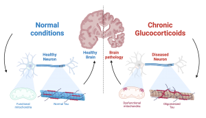
 First, Dr. Waites’ group established the role of chronic GC exposure in mitochondrial fitness and pathogenic Tau accumulation in vivo. Using the widely used synthetic glucocorticoid dexamethasone (DEX), they demonstrated that GC induce mitochondrial dysfunction (increased permeability and lower respiratory capacity) and oligomeric Tau accumulation. Next, Drs. Du and Yu very cleverly employed in vitro experiments to demonstrate that DEX triggered mitochondrial dysfunction by increasing mitochondrial permeability: their research showed that GC exposure stimulates the opening of the mitochondrial permeability transition pore, a big, non-discriminant, pathological hole in the mitochondrial membrane that when open, greatly compromises its function. Most importantly, they found that GC triggers opening of the pore by activating its key component, the protein cyclophilin D. This key finding led Dr. Waites’ group to block cyclophilin D activation, either genetically (i.e. changing the DNA of the protein) or using drugs, with great success: cells where cyclophilin D activation was prevented were remarkably resilient to GC stress, and showed much lower levels of mitochondrial dysfunction and pathological Tau accumulation.

 Although this was an extremely exciting result by itself, it posed a problem moving forward: neither genetical nor pharmacological inhibition of cyclophilin D are currently viable therapeutic approaches, due to their poor bio-availability or invasive methods required to deliver the treatments. So what other approach could they use to also prevent mitochondrial pore opening, while still being clinically tractable? After some digging, Drs. Du and Yu found apo-cyanin, an orally-available inhibitor of mitochondrial respiration that could tackle GC-driven brain pathology while not directly targeting cyclophilin D. In line with their predictions, this drug was able to restore mitochondrial function and prevent Tau pathology in DEX-treated cells. Most importantly, they were also able to restore neuronal health, neuronal connectivity, and prevent anxious and depressive behavior associated with GC treatment.

 All of these experiments clearly demonstrated that GC-exposure triggers mitochondrial dysfunction and Tau accumulation, which could in turn promote the development of brain pathologies. How could they demonstrate, however, that targeting mitochondrial permeability is a feasible approach to treat common brain pathologies such as Alzheimer’s disease? To tackle this question, Dr Waites’ group made use of an ingenious technique, where they replaced all mitochondria in a test cell with those coming from an Alzheimer’s disease patient. Excitingly, in this model, treatment with mito-apocynin was sufficient to partially reverse mitochondrial dysfunction and Tau oligomerization found in the cells containing mitochondria from Alzheimer’s patients.

 Dr Waites’ group story is a hallmark out-of-the-box thinking, and clinical relevance. While their research clearly highlights mitochondrial damage induced by GC and cyclophilin D activation as a key trigger of Tau accumulation, the implications of this finding could be much broader:  How generalizable is this mechanism to other forms of brain disease such as ischemia, inflammation, or aging? As a matter of fact, cyclophilin D levels are elevated in Alzheimer’s patients, and its inhibition is protective in mouse models of sclerosis, Parkinson’s, and Alzheimer’s disease.

While this story provided incredible insight into the role of mitochondria in controlling brain pathology, many other questions remain unanswered.  For example, What is the exact mechanism by which mitochondrial dysfunction promotes pathogenic Tau accumulation? It is highly likely that these two events are intertwined, and that oligomeric Tau can also drive mitochondrial dysfunction.

 Despite the still-unknowns, it is clear that after this publication from Dr Waite’s lab, we should re-contextualize our middle-school textbooks: The mitochondria is the powerhouse of the cell, and during stress, the gate-keeper of our brain’s health.

Reviewed by: Trang Nguyen, Erin Cullen 

Maternal Stress and the Developing Brain

As humans, we all experience stress. It is a normal, and sometimes even beneficial, part of life. A small amount of stress can help motivate someone to prepare for a job interview or study for an important exam. There are times, however, when stressors become too overwhelming and even detrimental to health. Scientists, from medical researchers to psychologists, have studied stress for decades and documented some of these negative impacts on the brain. When thinking about the importance of the foundational, early years of a person, the presence or lack of stress can play a crucial role in development. For instance, extensive research shows that living in poverty is extraordinarily stressful for families and can negatively influence children’s brain development. The impacts of stress resulting from situations such as growing up in poverty warrant further investigation, especially considering that in 2020, one in six children in the U.S. was living in poverty.

Researchers can use various methods to assess how factors like stress impact the brain of growing children. Developmental scientists can use a tool called EEG, short for electroencephalography, to study the brain. EEG measures electrical activity in the brain by recording the communication between brain cells. It is an ideal neuroimaging method for understanding infant brain development since it allows for infants to be awake and moving, and even sitting on their caregiver’s lap during recording. Besides being infant-friendly, EEG is a useful tool for looking at brain development, given that there is a known pattern of how brain activity changes across the first few years of life.

Specifically, when using EEG to look at brain development, scientists typically see two different patterns. Broadly, infants have a mix of different types of brain activity that we call low-frequency and high-frequency power. Low-frequency power (e.g., theta) tends to be higher when the brain is at rest, while high-frequency power (e.g., alpha, beta, and gamma) tends to be used for more complex thinking like reasoning or language. As infants grow, scientists see that low-frequency power decreases and high-frequency power increases. Importantly, we can use EEG to assess how factors like stress impact the tradeoff of low-frequency and high-frequency power in the developing brain.

Image of a one-month-old infant with an EEG cap.
Figure 1. A one-month-old infant with an EEG cap. Courtesy of the Neurocognition, Early Experience and Development Lab.

Research shows that children growing up in chronically stressful environments often show alterations in the typical pattern of brain activity development. To further understand the mechanisms underlying this pattern of development, scientists have begun to study which biological and environmental factors may be at play. For instance, researchers can examine the role of caregiver stress, socioeconomic status, home environment, and neighborhood factors, just to name a few.

A recent paper by Dr. Sonya V. Troller-Renfree and colleagues examined maternal stress by looking at the amount of stress hormone (cortisol) found in hair. This measure assesses chronic stress and provides researchers with the average cortisol level of the mother from the preceding 3 months. Dr. Troller-Renfree’s research group hypothesized that infants who have mothers with higher stress hormone, compared to mothers with lower levels of stress, would show differences in their brain activity. Specifically, the researchers predicted that infants of more chronically stressed mothers would exhibit proportionally more low-frequency power and proportionally less high-frequency power compared to infants with physiologically less-stressed mothers.

Indeed, their results showed that infants of mothers who had higher levels of hair cortisol demonstrated higher levels of low-frequency (theta) activity and lower levels of high-frequency (alpha and gamma) brain activity. This finding is consistent with previous research showing that stress and adversity impacts early neural development. Importantly, Dr. Troller-Renfree’s team sampled a diverse pool of participants (both in terms of socioeconomic status and race), therefore bolstering the generalizability of their findings.

So what are the implications of these alterations? Research suggests that similar patterns of neural activity are associated with negative outcomes later in a child’s life, including language development and psychiatric problems. Nevertheless, this does not mean that a child will undoubtedly experience these issues. Additionally, it may be possible that these patterns, while associated with negative outcomes in some areas, may also be adaptive in other circumstances. Furthermore, the issue of the mechanisms by which a mother’s stress impacts the developing child still remains unclear. How exactly does a mother’s stress level impact the brain of her child?

Based on previous research by other scientists, Dr. Troller-Renfree posits a few mechanisms that must be further explored. For example, it is possible that stress impacts crucial mother-child interactions. It could be that stress hormones are passed from mother to baby in utero or through breastmilk. Moreover, it is also possible that environmental factors impact stress and brain development.

It is crucial that developmental scientists continue studying these mechanisms so that targeted intervention programs can be formed for families facing stress. Indeed, the esteemed pediatrician and researcher Dr. Jack Shonkoff of the Center on the Developing Child said in an episode of The Brain Architects Podcast: “In fact, one of the cardinal principles of the science of early childhood development is that if we want to create the best kind of environment for learning and healthy development for young children, we have to make sure that the adults who care for them are having their needs met as well.” As a society, we must recognize how detrimental stress can be to the developing child and invest in finding effective ways to alleviate caregiver stress.

Dr. Sonya V. Troller-Renfree is a Goldberg Postdoctoral Fellow in the Neurocognition, Early Experience and Development Lab at Teachers College, Columbia University. Her research focuses on the effects of early adversity and poverty on cognitive and neural development. She intends to continue examining these questions as part of her new, federally-funded Pathway to Independence Award (K99/00). You can stay up-to-date on her research findings on Twitter at @STRscience or on her website: www.sonyatrollerrenfree.com.

Follow this blog

Get every new post delivered right to your inbox.