How a molecular structure explains the transport of fatty acids past the blood-brain barrier

The brain and eyes develop through constant circulation of nutrients through the blood-brain and blood-retina barriers. One such nutrient that is essential for development is an omega-3 fatty acid called docosahexaenoic acid (DHA). DHA makes up a fifth of all the fatty acids required on the membranes of cells in the central nervous system. Neither the neurons in the brain nor the cells in the eye are capable of synthesizing DHA by themselves and therefore depend on dietary sources for DNA. Previously, scientists knew from cellular clues that this fatty acid most likely passed through to the blood-brain and blood-retina barriers in the form of lysophophatidylcholine (LPC-DHA) using a molecular channel. This transporter is known as a major facilitator superfamily domain containing 2A, or MFSD2A, with the help of sodium atoms regulating the channel. However, it was not clear how this channel allowed the passing of complex molecules like DHA. A recent study by Dr. Rosemary Cater and colleagues at Columbia University provided precise clues to further show the structure of this channel. 

To investigate the structure of MFSD2A, the authors used a state of the art imaging technique called single-particle cryo-electron microscopy. This is a method of electron microscopy where a beam of electrons is transmitted through a rapidly frozen purified molecule. Because the sample is flash frozen, the molecules trapped in a frozen state can be imaged in their native shape as present in the cell and from multiple angles. By capturing and combining multiple captured 2D images, a 3D structure of the protein can be reconstructed with extreme accuracy. Cryo-electron microscopy is so impactful in biological significance that this method was awarded the 2017 Nobel Prize in Chemistry. found a number of molecular patterns and arrangements of protein chains that make up a full molecule of MFSD2A

Protein structure studies are typically among the most challenging grounds to explore in biology because proteins need to be captured in their native state as present in the cell. Past discoveries of various protein structures have been so instrumental in shaping therapeutic areas that the extent of mechanistic understanding of biological molecules has resulted in recognition by Nobel committees. Most recently, the discovery of the structure of the ribosome opened up fields of exploration into therapeutic interventions into ribosome diseases, some of which can lead to cancer.

To get the best chance at imaging the structure of MFSD2A, the scientists extracted and examined purified versions of this protein obtained from multiple organisms: the zebrafish, the frog, European cattle, the domestic dog, the red junglefowl, mice and humans. Finally, the authors found that the protein obtained from the red junglefowl, which is a rooster species that originates from Southeast Asia, was the most experimentally stable and most alike (73% identity) the human version of MFSD2A. 

Using additional accessory proteins to help with the orientation of MFSD2A, the authors obtained high-quality images, with a resolution of 0.3 nanometer, or 0.3 billionth of a meter. From the imaging data, the authors found that MFSD2A protein itself is about 5 nm wide and 8 nm long. MFSD2A is a transporter protein and like many transporters, it contains repeated bundles of helices made of protein chains that traverse the cell membrane and are connected by a protein chain that loops within the space in the cell. 
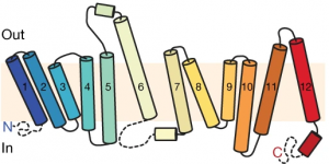
Structure of MFSD2A arranged as protein helices (colored cylinders) within the cell membrane along with protein loops that form both in the extracellular space (“Out”) and within the interior, cytoplasmic space (“In”) of the cell. The cytoplasmic loops likely have an important functional role. Figure from Cater et al, 2021.

The cell membrane consists of two layers of lipid molecules, known as the lipid bilayer, that allow entry and exit of materials from the cell. These loops provide the shape to the protein inside the cell such that it appears to provide a large enough cavity opening from the lipid bilayer into the cellular space to allow the target molecules to enter the cell. Amino acids are the building blocks of proteins and the cavity contains amino acids of both water-attracting and water-repelling kind. This property makes it possible for many molecules of differing chemical nature to be able to be accommodated within the cavity. This cavity contains three important regions that allow for the protein to be specific and functional: a charged region, a binding site for sodium atoms and a lipid-specific pocket. The authors speculate that these parts help in establishing the mechanism by which LPC-DHA is transported from the outside into the cell. The multiple protein helices form two protein domains that capture LPC-DHA from outside the cell layer of the blood brain barrier of endothelial cells, then rock over a rotation axis so that now their confirmation switches and finally, they release the protein molecule into the cell. For this activity of movement of LPC-DHA, sodium atoms are absolutely required to allow for the shape change of the protein. Once LPC-DHA enters the barrier cells in this manner, the protein is then transported across to the other side of the cell facing the brain containing neurons. 
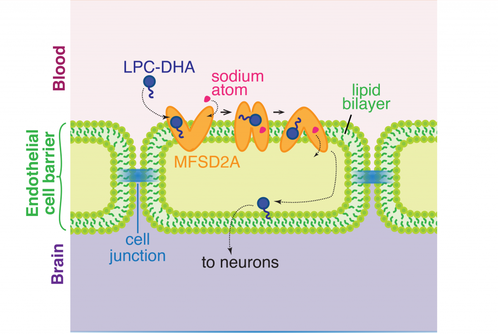
The transporter channel MFSD2A changes its shape once it binds sodium atoms in the extracellular space, which helps the transport of LPC-DHA from the blood into the brain space through the barrier of a single line of cells made up of endothelial cells. Figure adapted from Cater et al, 2021.

Humans with mutations in MFSD2A gene have abnormal brain defects such as microcephaly, and disruption of the gene in mice affected neuronal branching and fatty acid composition in the brain. The discovery of the structure of a molecule that mediates uptake of essential nutrients across the blood-brain and eye-brain barriers will help in the delivery of therapies of neurological diseases.

Dr. Rosemary J. Cater is a postdoctoral researcher in the lab of Dr. Filippo Mancia in the Department of Physiology and Cellular Biophysics at Columbia University.

Magic under the microscope

Researchers design an accessible, straightforward technique to characterize moiré systems – a class of materials built by placing slightly misaligned atomic monolayers on top of each other. Under certain conditions, such moiré structures exhibit exotic physical phenomena absent in the individual units that conform them.

A moiré pattern is an interference effect that arises when two grids are superimposed. It can be observed in the wrinkles of a mesh shirt and it is responsible for the fringes that appear when taking a picture of a computer screen. Moiré patterns are present in art and fashion, and in the last few years their effect in two-dimensional materials has entailed a revolution in physics.

Two-dimensional materials are those that are less than a nanometer thick. The first one to be isolated was graphene, a single-layer of carbon atoms (see Fig. 1a). Such discovery opened a whole new field of research and many labs around the world started making their own stacks – structures with two-dimensional materials placed on top of each other. If one were to place one of those layers slightly misaligned with the one below, a moiré pattern would emerge. This interference effect can be visualized in Fig. 1b. The small circles represent the carbon atoms that form a crystalline lattice (an ordered structure) on each graphene layer. The top layer is rotated with respect to the bottom one and, as a consequence, a periodicity larger than the atomic lattice emerges as highlighted in Fig. 1b.

In 2018, the field of condensed matter physics was stirred up: such moiré materials, at a very specific misalignment value called the magic angle, exhibit electronic states of matter that are not present in the individual layers, such as superconductivity or magnetism. The emergence of those electronic phases is a consequence of the moiré pattern and its direct visualization is critical for their understanding. There are a few techniques, including transmission electron microscopy and scanning tunneling microscopy that allow for this, but they require complex setups that do not necessarily work for any material, which has significantly slowed down the progress in the field. McGilly and colleagues show a new and simple technique based on piezoresponse force microscopy to visualize moiré patterns.

A piezoresponse force microscope consists of a sharp metallic tip brought into contact with the material under study –  in this case, the moiré system (see Fig. 1c). Piezoresponsive materials are those that undergo a mechanical deformation in the presence of an electric field. In the microscope, the sample moves a small amount when a voltage is applied across it and the tip follows the motion. Such tip motion is measured as a voltage which is amplified to detectable values. The tip is then moved around the sample and the process is repeated on every pixel of a selected region, producing a map of the sample’s deformation.

a. Graphene atomic lattice. Each ball represents a carbon atom. b. Twisted graphene bilayers. The three main stacking configurations are shown (AA, AB and domain wall). The moiré unit cell is highlighted. c. Microscope tip in contact with the graphene bilayer d. The strain on the graphene layer bends the chemical bonds between the atoms from in-plane (left) to a mixed in-plane/out-of-plane character (right).

In principle, it was not obvious that a moiré pattern would be detectable with the microscope. When moiré patterns form, it creates a repetitive set of individual units that are called unit cells (highlighted region in Fig. 1b). Each unit cell is formed by regions with different atomic three-dimensional configurations, called sites. In the case of graphene, those sites are called AA and AB which stands for how the atoms from each layer lie on top of each other (see insets in Fig. 1b). The AB regions (also called domains) are separated by domain walls, as highlighted in Fig. 1b. McGilly and colleagues show that the voltage signal detected with the microscope is localized on the domain walls.

When the moiré pattern forms, the atomic layers relax to accommodate it and the layer wrinkles along the domain wall (see right panel in Fig. 1d). Since the microscope is not sensitive to such small deformation, the origin of the detected signal must be electronic. Flat graphene layers have planar bonds, as shown in the left panel of Fig. 1d. However, the curvature of the wrinkle bends the atomic bonds on the graphene layer, which in turn causes an asymmetric distribution of the charge in the vertical direction and gives rise to an out-of-plane polarization (P), which is responsible for the signal measured in the microscope.

The technique designed by McGilly and colleagues has been proven extremely useful for the advancement of the field due to the simplicity of the method and the fact that it allows imaging of any moiré pattern, independently of the nature of the individual units that conform it – that is, whether they are metals, semiconductors or insulators. Being able to image moiré patterns with such an accessible technique will help improve the fabrication process, and having uniform samples is critical since strain gradients can significantly alter the states of matter that emerge in moiré materials.


Dr. Leo McGilly is a Postdoctoral Research Fellow in the Physics Department at Columbia University.

Follow this blog

Get every new post delivered right to your inbox.