For most people, the famous words “Who wants to live forever?” by the British rock band Queen seem merely hypothetical. However, scientists have been trying to identify the secret of immortality for decades. Their research has revealed an important role of the biological clock in regulating lifespan. The biological clock is a natural timing device composed of small molecular “clocks” in cells throughout the body that together dictate circadian rhythm, a term that originates from the Latin words circa (around) and dies (day). As the name implies, circadian rhythm refers to all natural processes that have a period of roughly a day, such as the sleep/wake cycle, body temperature change, and release of hormones like melatonin and cortisol. Because organisms, including humans, lose circadian rhythmicity with age, scientists thought that loss of circadian regulation contributes to aging and limits lifespan. However, recent findings by Dr. Matt Ulgherait and colleagues from the Department of Genetics and Development at Columbia University, show that the relationship between circadian rhythm and lifespan is more complex than initially thought.
Dr. Ulgherait studied the role of genes that regulate cell-intrinsic rhythms, so called “clock genes” in aging. To this end, he used the model organism Drosophila, also known as the fruit fly. Although the evolutionary distance between fruit flies and humans is large, they show a remarkably high degree of genetic similarity: about 75% of the disease-causing genes in humans match up with the genome of Drosophila. In addition, the relatively short lifespan of fruit flies of about 50 days makes them a very practical model for aging research. Dr. Ulgherait introduced loss-of-function mutations in four different clock genes in the flies, named “cycle”, “period”, “timeless”, and “clock”, and found that only disruption of cycle and clock decreased lifespan, while disruption of timeless and period surprisingly extended lifespan by about 15-20%.
The researchers continued by investigating the specific role of period, named for its contribution to the length of circadian cycles, to find out how this gene negatively affects lifespan. Dr. Ulgherait observed that period mutant flies not only lived longer than their genetically intact counterparts, but were also leaner despite an increased food intake. Remarkably, nutrients that were taken up by the flies were not converted into storable energy but rather used for heat production, as reflected by a higher ability of period mutant flies to recover after a cold shock of 4 °C for 1 hour. When burning of nutrients is disconnected from energy production, the metabolic machinery in the cell is considered to be “uncoupled”, a process regulated by so-called “uncoupling proteins”. Dr. Ulgherait found that the expression of uncoupling proteins was consistently high in period mutant flies. Moreover, disruption of uncoupling proteins reverted lifespan of period mutants to that of control flies, indicating that uncoupled energy metabolism and increased heat generation is important for longevity.
To determine which organ of the body is responsible for the effect of period on aging, the researchers removed the gene from different tissues one by one. This way, they found that loss of the period gene in the intestine was sufficient to increase lifespan. Intestinal expression of uncoupling proteins was required for the increased lifespan in period mutant flies, indicating that an uncoupled energy metabolism in the gut is essential for longevity. To understand the underlying mechanism through which uncoupled energy metabolism in the intestine regulates lifespan, Dr. Ulgherait examined intestinal functions that are affected by aging, including intestinal barrier function, which deteriorates with aging and makes the intestines more leaky. The scientists assessed intestinal barrier function in period mutant flies by performing the “smurf assay”. This assay, named after the children’s cartoon, measures leakage of an ingested blue dye which makes the fly resemble a smurf (see image below). Indeed, period mutant flies showed a lower percentage of “smurfs” relative to controls, indicating less intestinal leakiness. Thus, loss of the circadian period gene protects against aging-related intestinal dysfunction.
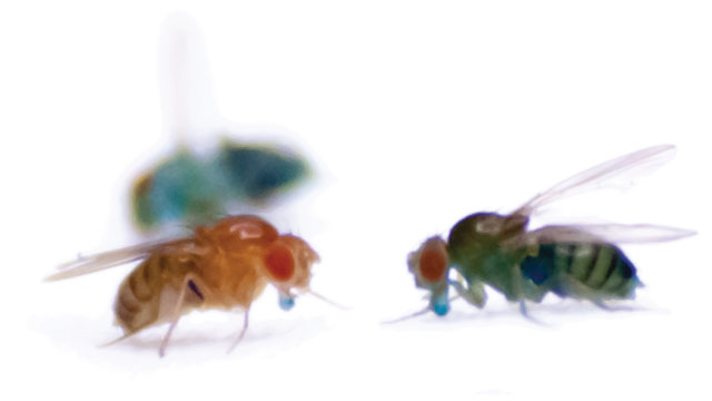
Photograph showing a normal-colored fruit fly (bottom left) with an intact intestinal barrier function and “smurf” flies (right and top left) with a disrupted intestinal barrier function. Source: The Scientist.
In summary, the research by Dr. Ulgherait and colleagues shows that disruption of circadian rhythm affects lifespan by modulating uncoupled energy metabolism in the gut. Although this research was performed in Drosophila, genetic variability in uncoupling proteins has been shown to predict longevity in humans. Therefore, pharmacological targeting of uncoupling may be one of the keys for increasing lifespan. So perhaps we should avoid hypotheticals and actually start asking the question: “Would you want to live forever?”