How many candles on the cake this year?

Every year as we celebrate our birthdays, we mark the addition of a year to our lives. Our birthdays determine our chronological age measured in days, months and years since the day we were born. Biological aging on the other hand is another measure of aging that accounts for the gradual accumulation of cellular and tissue damage that occurs in the body as we grow older. Aging is a natural process and various factors contribute to biological aging, including our chronological age, genetics, lifestyle, nutrition, and physical activity. Research has shown poor nutrition and low physical activity can accelerate biological aging. Accelerated biological aging is marked by increased levels of certain hallmarks of cellular damage, leading to chronic diseases. Poor nutritional habits and sedentary lifestyles have been associated with increased risk of heart diseases, high blood pressure, cholesterol and type 2 diabetes. Additionally, over 60% of the aging population (>65 years) is expected to be affected by more than one chronic disease by 2030. Research has also shown that lifestyle interventions may reduce or delay the progress of biological aging. In this regard, Aline Thomas and colleagues obtained real life data from a large cohort of US adults to study the association between lifestyle behaviors and biological aging using mathematical models. They assessed signs of aging in individuals who engaged in some form of moderate to vigorous physical activity in their leisure time and followed a diet that resembled a mediterranean diet compared to individuals who followed a less-healthy lifestyle. A Mediterranean diet focuses on plant-based foods and healthy fats. It includes vegetables, fruits, whole grains, fish and extra virgin olive oil as a source of healthy fats. The researchers studied diet, exercise, and variations in healthy lifestyle behaviors across different age groups, genders, and body mass indices (BMI).

Dr. Thomas and colleagues combined data collected over a period of 20 years from 1999-2018 for their study. The study included 42,625 participants between the ages of 20-85 and assessed the adherence to the Mediterranean diet and an exercise regimen using a point based system. Inclusion of fruits and vegetables, legumes, cereals, fish and a ratio of mono-unsaturated to saturated fats were each awarded one point. A healthy Mediterranean diet also includes a mild-moderate amount of alcohol, which is 0-1 glass for women and 0-2 glasses for men. So, a point was given if a mild-moderate amount of alcohol was consumed. Dairy products and meat are not part of the Mediterranean diet. If participants had consumed these foods but had consumed it less than a specific amount, they were still awarded a point. The points were totaled and found to be between 0 and 9. Higher scores meant a higher adherence to the Mediterranean diet. Leisure time physical activity (LTPA) describes any physical activity performed during participants free disposable time. The researchers assessed LTPA based on the frequency, duration and intensity to calculate points / scores for each activity. They categorized the activity levels based on the scores per week into four groups ranging from – sedentary (0 points), low (<500 points), moderate (500-1000 points) and high (>1000 points). Biological age was calculated using an algorithm called PhenoAge. The algorithm calculates biological age based on chronological age and 8 biomarkers obtained from blood samples.

The study included individuals across different races, socio-economic backgrounds, marital statuses, income to poverty ratios, and with various lifestyle-related factors (e.g., smoking, BMI category, total energy intake), making it a representative population of US adults. The researchers found very interesting observations relating to diet and exercise. They discovered that adherence to a relatively healthy diet and engagement in  physical activity were independently associated with a lower biological age. Participants with a healthy diet and some level of activity were on average 1 biological year younger than the participants with the least healthy diet and sedentary lifestyle. Another very interesting finding was that individuals who had a less healthy diet but who were active even at a low level showed delayed biological aging. However, delayed biological aging was not found in participants with a healthy diet and a sedentary lifestyle, suggesting that moderate physical activity is a key component of healthy biological aging.

The findings of this study reiterates the need for better lifestyle choices across all strata of the population as the results were consistent regardless of age, sex and BMI category. A nutritious diet and moderately active lifestyle can have a positive impact on health, aging and quality of life. Getting older is inevitable, but you may be one year younger with a healthy diet and an exercise routine.

Reviewed by: Trang Nguyen, Giulia Mezzadri, Erin Cullen, Maaike Schilperoort 
 

A pharmacological approach to lifestyle related metabolic disorders- does it make anti-sense?

Non-Alcoholic fatty liver disease (NAFLD) is a group of liver diseases that include NAFL and Non-alcoholic steatohepatitis (NASH). It is a chronic progressive disease of the liver not caused by alcohol. It starts with fat accumulation, progresses into inflammation, swelling and liver enlargement (NASH), fibrosis, cell death and replacement of dead cells by scar tissue(cirrhosis) and finally results in cancer (hepatocellular carcinoma). A figure describing this progression has been published by my colleague Maaike Schilperoort in an article describing emerging therapeutic strategies for fatty liver-related cancer. 

NASH is the most severe form of NAFLD before progressing into the irreversible stages of cirrhosis and cancer. It remains under-diagnosed as it is asymptomatic, or it is accompanied by non-specific symptoms. Individuals with hypertension, high cholesterol, who are over-weight or obese, have diabetes or insulin resistance are at a greater risk to develop NASH. It is largely a lifestyle associated metabolic disorder made worse in individuals with obesity and type 2 diabetes. Current treatment modalities focus on lifestyle interventions and management of co-existing conditions. A lack of specific and targeted pharmacological recommendations with proven efficacy complicates NAFLD management.

Junjie Yu and colleagues have conducted a comprehensive study by using data available from a clinical trial of patients with NASH and studying mouse models of NASH. They identified a gene – Jagged 1 (JAG1) that was increased in patients who had NASH and fibrosis. They used this key finding to conduct experiments on mouse models to further study the role of JAG1 in either reducing or worsening NASH and liver fibrosis. They have also used a cell targeted strategy to test potential therapeutic interventions associated with the development of NASH. 

To mimic human NASH, they fed mice with a NASH inducing diet, rich in saturated fat, sucrose, and cholesterol as well as fructose containing drinking water. The mice develop liver steatosis, inflammation, fibrosis, weight gain and insulin resistance which are symptoms seen in patients with NASH. JAG1 was increased in the liver, and this correlated with an increase in fibrotic markers in mice fed a NASH-inducing diet. An interesting observation that directed the rest of the study was that JAG1 was increased in the liver specific cell type called hepatocytes. They used a virus mediated gene delivery method to increase or decrease Jag1 in the hepatocytes of the mice that were fed the NASH-inducing diet. Increasing Jag1 increased fibrosis that is induced by the NASH diet in the mice and decreasing Jag1 protected the mice liver from developing fibrosis. Based on these insights they used a technology called antisense oligonucleotides (ASO) to block Jag1 expression in mice that were fed a NASH-inducing diet. They found mice treated with Jag1-ASO had reduced expression of JAG1 at the gene and protein levels along with a reduction in inflammatory and fibrotic markers. However, as this method would target all cell types, a hepatocyte specific Jag1 inhibitor was developed. Mice fed with NASH-inducing diet on treatment with hepatocyte specific Jag1 inhibition show decreased Jag1 in the liver as well as reduction in liver fibrosis.

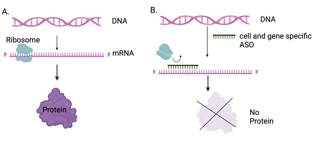
This is a very interesting approach that could lead to specific and targeted pharmacological treatment of NASH. ASOs are short single strand nucleotide sequences that can be produced to target specific genes of interest (like Jag1 in this case) in cells. They alter protein expression during the process of translation from RNA to protein (Fig 1). As they are made to target specific genes and cells, they have a higher chance of success. Currently there are 15 FDA approved ASO based drugs for disorders ranging from neurodegenerative disorders to cancer. The main limitation of ASO is the enzymatic degradation of oligonucleotides and removal from the body by the kidneys.  Further research into improving ASO to optimize delivery and safety could lead to development of therapies for disorders that require targeted pharmacological interventions.

Figure 1. A. A schematic of regular transcription and translation processes involved in protein synthesis. B. ASO mediated disruption of protein synthesis. Figure created using Biorender.com

Currently, pharmacological therapies for NAFLDs are recommended for individuals who do not achieve expected weight loss and for those individuals with stage 2 or greater NASH-induced fibrosis. Lifestyle changes may not be possible for all individuals with metabolic disorders due to various reasons including socio-economic reasons, limited food resources, disabilities, etc. Though lifestyle interventions like weight management, maintaining a healthy diet and regular exercise have been shown to reduce symptoms and manage the disorder, it works well if it is diagnosed at earlier stages. However, given that NASH does not have specific symptoms and is grossly under-diagnosed, an option for treatment of the disorder when it is at later stages may alleviate the disease burden. Specific targeted pharmacological approaches towards treating a metabolic disorder would be a feasible approach and may be a more efficient way to treat NAFLD when combined with lifestyle changes.

Reviewed by : Trang Nguyen and Samantha Rossano


“CRACK”ing cocaine addiction with medication

Cocaine is a highly addictive stimulant drug made from the leaves of the coca plant that alters mood, perception, and consciousness. It is consumed by smoking, injecting or snorting. According to the United Nations Office on Drugs and Crime, an estimated 20 million people have used cocaine in 2019, almost 2 million more than the previous year. Cocaine causes an increase in the accumulation of dopamine in the brain, which is a chemical messenger that plays an important role in how we feel pleasure and encourages us to repeat pleasurable activities. This dopamine rush causes people to continue using the drug despite the cognitive, behavioral and physical problems it causes,leading to a condition referred to as cocaine use disorder (CUD). CUD related physical and mental health issues range from cardiovascular diseases like heart attack, stroke, hypertension, and atherosclerosis, to psychiatric disorders and sexually transmitted infections. 

According to the CDC, cocaine use was responsible for 1 in 5 overdose deaths. Though almost all users who seek treatment for CUD are given psychosocial interventions like counseling, most continue to use cocaine. Pharmaceutical medication may increase the effectiveness of psychosocial interventions. Medications for other substance abuse disorders (opiod and alcohol) have shown to block euphoric effects, alleviate cravings and stabilize brain chemistry. However, there are currently no FDA approved drugs to treat CUD. 

Dr. Laura Brandt and colleagues have systematically reviewed available research up until 2020 in the area of pharmacological CUD treatment. In this review, they discuss the potential benefits and shortcomings of current pharmacological approaches for CUD treatment and highlight plausible avenues and critical considerations for future study. The authors reviewed clinical trials where the primary disorder is cocaine use and medication tested falls into four categories: dopamine agonists, dopamine antagonists/blockers, new mechanisms that are being tested and a combination of medications. 

Dopamine agonists are medications that have a similar mechanism of action as cocaine, i.e., they can act as substitutes for cocaine without the potential adverse health effects. Dopamine releasers and uptake inhibitors fall under this category and have shown the most promising signs thus far for reduced cocaine self-administration in cocaine-dependent participants. Dopamine uptake inhibitors bind to the dopamine transporter and prevents dopamine reuptake from the extracellular space into the brain cell. The medications that act as substitutes result in the users exhibiting blunted dopamine effects such as low levels of dopamine release and reduced availability of dopamine receptors for dopamine to bind to. They help reduce dopamine hypoactivity by slow release of dopamine which in turn helps reduce responses such as cravings for cocaine and withdrawal symptoms which is usually a cause for relapse. A common concern associated with using dopamine agonists is the possibility of replacing cocaine addiction with the medication. However, there is no strong evidence for this secondary abuse as well as for the cardiovascular risk when using the agonist as a means of treatment. 

Dopamine antagonists/blockers are substances that bind to dopamine receptors, preventing the binding of dopamine and thereby blocking the euphoric effects of cocaine. This approach facilitates the decrease in cocaine use as the effects of cocaine use are absent. Antipsychotics medications, anti-cocaine vaccines, modulators of the reward system, and noradrenergic agents fall under this category. This approach is generally considered to be less effective in treatment for CUD as they require high levels of motivation to start the treatment as well as to maintain it. 

New medications are those that are currently in clinical trials and are being tested in humans for the treatment of CUD. Combination pharmacotheraphy is an interesting approach for treatment and involves combining two medications to treat CUD. An absence of FDA approved medications limits exploration in this direction. 

Having reviewed these data and their shortcomings, the authors point out a very important factor in these studies – the shortcomings of the studies depend on more than just the medication. On one hand, limitations due to medical procedures such as the dosage of medication and its formulation, completion of the medication course, providing/not providing incentives to participants of the study may have hindered the success of these studies. On the other , individuals seeking treatment are not all the same. They differ in terms of cocaine use severity, presence of mental health illness, substance use disorders apart from cocaine use, and their genetics may also play a role in the success of their treatment.  Pharmacotherapy formulations for CUD is not a one size fits all but needs to be tailored to the individual seeking treatment as well as the substance used.  A combination approach targeting withdrawal of the drug and allowing patients to benefit more from behavioral/psychosocial interventions would be more helpful on their path to recovery. Another very important point that requires some attention is the method for determination if the medication has worked. Most studies use the gold standard of performing qualitative urine screens to determine sustained abstinence in clinical trials of pharmacotherapies for CUD. Urine toxicology as evidence of treatment success is not a clear-cut method as various factors impact interpretation of the results. Second the medication is considered to successfully treat CUD only when there is complete abstinence from cocaine use. As many physical and psychological issues accompany substance abuse, considering CUD treatment to be linear is not very beneficial. Considering other aspects such as improvement in quality of life and ability to carry out daily activities would be a better indicator of the effectiveness of the medications used. 

With an increase in cocaine use and abuse in recent years, there is an urgent need to identify medications to treat CUD. The review consolidates the current approaches to treating CUD with medication and points out factors that are overlooked while interpreting the results from these studies. Tailoring medications to each individual would greatly improve clinical trial outcomes and have higher success rates for treating substance use disorders- a promising avenue that needs to be explored.

Dr. Laura Brandt is a Postdoctoral Research Fellow in the Division on Substance Use Disorders, New York State Psychiatric Institute and Department of Psychiatry Columbia University Irving Medical Center.

Reviewed by: Trang Nguyen, Maaike Schilperoort, Sam Rossano, Pei-Yin Shih


Beneath The Surface Of Healing Wounds

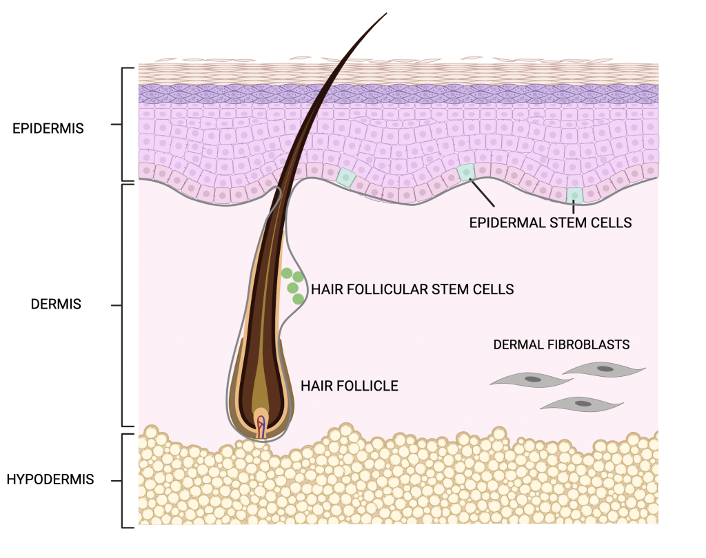
With an average weight of ~12 kg and surface area of ~2m², skin is the largest organ of the body and is made up of three layers: epidermis, dermis, and hypodermis. Epidermis, the outermost layer, is composed of densely packed epithelial cells. Under the epidermis lies the dermis, which mainly contains blood vessels, hair follicles and sweat glands. The third layer, the hypodermis, is composed of loose connective and fatty tissue (Fig. 1). Given its large area and exposure to external elements, the skin is susceptible to injury, ranging from minor bruises to cuts, lacerations, and tears. The skin’s response to insults like these is a process well-known as wound healing, which occurs in three stages: inflammation, proliferation, and tissue remodeling. These stages work together in an overlapping sequential manner to ensure complete wound healing. A failure in any of the normal wound healing stages leads to chronic wounding and aberrant scar formation as seen in burn injuries and scar tissue formations. A delay in wound healing can result in increased infections and permanent tissue damage as seen in patients with diabetes.

Fig.1. Schematic representation of the layers of skin.

During inflammation, there is an influx of immune cells to clear invading microbes and cell debris at the site of injury. This is followed by  the proliferation phase during which there is an increase in the production of epithelial cells  that will migrate to the outer edge of the site of injury to repair the wound and restore it back to its uninjured state.  An essential step in wound healing is the mobilization of stem cells for the formation of new epithelial cells. Skin stem cells are found in the basal layer of the epidermis and in the bulge area of hair follicles. Epidermal stem cells are actively involved in replenishing cells as skin undergoes normal homeostasis as well as during wound repair. Stem cells in the hair follicles, on the other hand have periodic patterns of rest and activity during hair growth. Following injury, however, hair follicular stem cells are also involved in rebuilding the epidermis to seal the open wound.

Interactions between the immune cells during the inflammation stage and stem cells during the proliferation stage of wound healing are important for efficient tissue repair to take place. Cytokines are signaling molecules produced by cells that are required for cell-cell communication to stimulate cell migration towards the site of injury. Molecular and cellular mechanisms to address the role of cytokines in mediating interactions between immune cells and stem cells during wound healing remain unexplained.

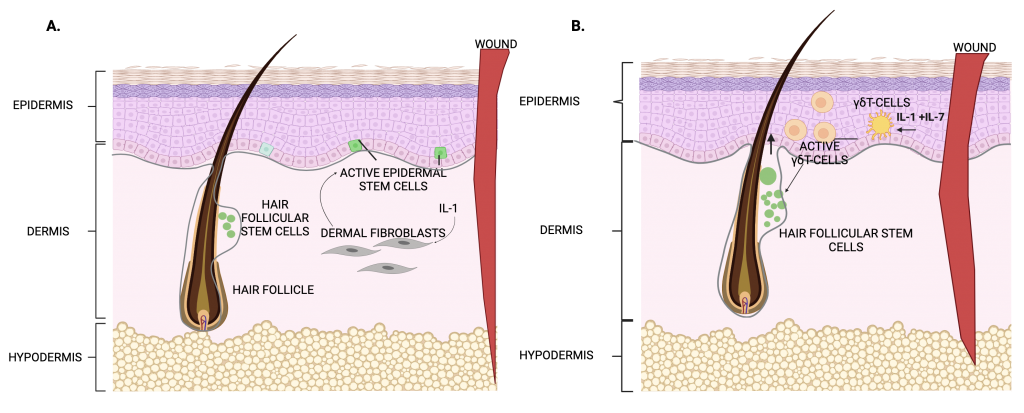
In a previous study, Pedro Lee and colleagues observed that mice that lacked the interleukin -1 receptor (IL-1R) for IL-1 cytokine had a delayed wound healing response. In the skin, IL-1 is released by damaged keratinocytes (keratin producing cells present in nails, hair and skin). and dysregulation of IL-1 has been associated with a number of skin diseases. Pedro Lee, Rupali Gund and colleagues conducted the current study to understand the mechanism behind delayed wound healing in IL-1R mutant mice. They analyzed the molecular and cellular interactions during wound healing using a genetically engineered mouse model in which the entire skin mimics the biological response of wound healing. This mouse model, which lacks the caspase 8 gene in the epidermis, exhibits a wound healing response even in the absence of injury, thereby providing the researchers with a large number of stem cells participating in a wound healing process. The authors studied the structure of tissues and gene expression patterns in the skin of these mice. The researcher additionally performed assays to analyze proliferation of cells by growing cells in the lab from the genetically engineered model and the mice lacking IL-1R. The researchers found that IL-1 mediates wound healing through activation of stem cell proliferation in two possible ways. The first is by activating dermal fibroblasts that will activate the epidermal stem cell to cover up the open wound (Fig.2.A). The second is the activation of a population of immune cells called gamma delta T(γδT)-cells. These cells in turn activate the resting stem cells found in the hair follicles. These activated stem cells then migrate from the hair follicle towards the site of the injury for wound healing. The researchers also found that IL-1 interacts with another cytokine, IL-7, and together they work to increase the number of active gamma delta T cells in wounded skin and secrete growth factors (Fig.2.B) thereby increasing the population of stem cells promoting wound healing.

Fig.2. Schematic of cytokine mediated interactions between cell types during wound healing. A. IL-1 mediated interactions between dermal fibroblasts and epidermal stem cells. B. IL-1 mediated interaction between immune cells (γδT) and stem cells. Image adapted from Lee, Gund et al., 2017

Normal wound healing is an important and complex physiological process to ensure timely healing to maintain skin integrity. It requires coordinated interactions between various factors, cells and cytokines at each healing stage. Lee, Gund and colleagues have identified a novel role for immune cells (γδT-cells) in tissue repair in addition to their well-established role of fighting infections. The ability of γδT cells to respond to IL-1 and, in turn, secrete growth factors that promote stem cell reparative activity expands the kind of functions that immune cells can perform in tissues in addition to their common role in immunity. Stem cell therapy and regenerative medicine shows great potential in the field of wound healing and skin regeneration.

This study reveals how immune cells communicate with stem cells during tissue repair and identifies new cellular interactions that can be targeted to prevent diseases in which wound healing is impaired. Such therapies will preclude need for invasive surgical interventions and skin graft procedures as a treatment for chronic wounds.

Dr. Rupali Gund is a postdoctoral research scientist in the department of dermatology at Columbia University Irving Medical Centre. Her research focuses on studying mechanisms in skin autoimmune diseases and finding new ways to design therapies to improve patient’s quality of life. 

Transcription factors and cellular fixer-uppers

Self-renewing stem cells are capable of developing into certain specialized cell types thus making them ideal candidates to study human development and as potential treatment modalities for a range of diseases. There are three types of stem cells: embryonic stem cells, adult stem cells and induced pluripotent stem cells. As the name suggests, embryonic stem cells are found in the embryo at very early stages of development. Adult stem cells are found in specific tissues post development. However, using human embryonic stem cells in research is quite restricted due to ethical, religious, and political reasons. This limitation has resulted in the identification of cell reprogramming techniques to convert differentiated cells, such as skin cells, back to an embryonic stem cell state through a process called induced pluripotency. The resulting induced pluripotent stem cells (iPSCs) are equivalent to the natural human embryonic stem cells and can be differentiated to any desired cell type using a mixture of biological molecules.

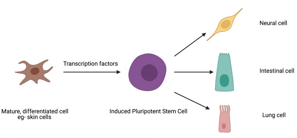
Cell reprogramming techniques can be likened to fixer-uppers. Imagine trying to remodel a building for a different purpose – converting an office building into a residential one for instance. Though the building material can be reused, with the aid of experts, there would be some structural changes and remodeling necessary to make it a home. Similarly, cellular reprogramming is the technique by which one cell type can be converted to another cell type in the lab with the help of certain gene expression regulators called transcription factors (Fig. 1). The process of inducing pluripotency has been studied extensively and the overexpression of four transcription factors – OCT4, SOX2, KLF4, cMYC (collectively referred to as “OSKM”) – has been shown to induce pluripotency in mouse skin cells.

Many studies have tried to identify other transcription factors with the potential to induce pluripotency or to replace OSKM in an effort to enhance the efficiency of iPSC generation. Of these four transcription factors, SOX2, KLF4 and cMYC have been successfully replaced by members of their protein family to induce pluripotency. However, replacing OCT4 with structurally similar and evolutionarily related factors failed to show similar reprogramming capabilities. This could indicate the presence of special molecular features on OCT4 that give it the ability to reprogram cells. However, these special features and the molecular mechanisms that enable OCT4 to induce pluripotency remain to be identified.

Fig.1. Depiction of pluripotency induction in differentiated cells. Transcription factors regulate the process of converting a mature cell into an induced pluripotent stem cell which can then be directed to differentiate into any desired cell type. Illustration created with BioRender.com

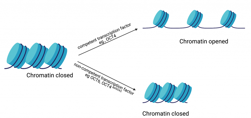
In the current study, Dr. Malik and colleagues hypothesized that the ability of a transcription factor to reconfigure chromatin (the complex of macromolecules composed of DNA, RNA, and protein, which is found inside the nucleus of eukaryotic cells), is one of the features that distinguishes a reprogramming competent transcription factor from a non-competent one (Fig. 2). To test this hypothesis, they studied the well-established OCT4-SOX2 relationship from initiation to maintenance of pluripotency. They performed their study by comparing DNA-accessibility, DNA-binding,  and transcriptional control by OCT4, OCT6 and an OCT4 mutant that does not interact with SOX2 (OCT4defSOX2) during early, mid and late phases of cell reprogramming. What makes this study particularly interesting is the fact that a previous study by the same group has shown that OCT4 naturally interacts with SOX2 to induce pluripotency, whereas OCT6 can only induce pluripotency when OCT6 was mutated to enhance its interaction with SOX2. Dr. Malik’s current study focuses on the mechanisms by which the above-mentioned transcription factors interact with chromatin and in turn bind to the transcription factor binding sites on the genes that are involved in processes from the initiation to maintenance of induced pluripotency.

Fig. 2. Depiction of chromatin remodeling by competent vs non-competent transcription factors. Opening up the chromatin by competent transcription factors and making transcription factor binding sites accessible is required to induce pluripotency. Failure to do so by non-competent transcription factors results in a failure to induce pluripotency. Illustration created with BioRender.com.

From this study, the researchers found that OCT4, OCT6 and OCT4defSOX2 have unique transcription factor binding sites on the pluripotency-related genes which could explain why substituting OCT4 with related transcription factors does not activate these genes. The results from this study challenge previously established roles for OCT4 in driving pluripotency. Dr. Malik and colleagues have identified distinct modes of chromatin interaction and roles for SOX2 and OCT4 during initiation, progression and maintenance of pluripotency. They found SOX2 to be a better facilitator of chromatin opening and initiator of pluripotency compared to OCT4. Once the cells have been initiated towards pluripotency, OCT4-SOX2 binding is required to see the process through and once the cells are pluripotent OCT4-SOX2 binding becomes less essential. The most important role of OCT4, they found, was to maintain the cells in a pluripotent state as opposed to its previously investigated role as an initiator of pluripotency. 

The results from this study contribute new insights to a rapidly progressing field. Identifying the roles of key factors during the stages of reprogramming would add vital pieces of information to the big puzzle of cellular reprogramming. These pieces of information would considerably enhance the use of stem cells as potential therapeutic candidates for a number of diseases .

Dr. Vikas Malik is a Postdoctoral Research Fellow in Dr. Jianlong Wang’s lab in the Department of Medicine at Columbia University Medical Center and is a member of CUPS and the Outreach and Communications Committee.


Follow this blog

Get every new post delivered right to your inbox.