What’s in Your Water? Arsenic Contamination and Inequality.

Water is one of the most essential elements for life. Every living creature requires access to a water source, humans being no exception. Unfortunately, access to clean drinking water continues to be a challenge for many individuals across the globe.  Systematic studies of water inequalities in the U.S. alone indicate increased contamination in areas often dismissed or underserved. Arsenic, a human carcinogen, or cancer causing substance, predominantly released from water flowing through rock formations has previously been measured at dangerous levels in  U.S. water sources. This finding led to the U.S. Environmental Protection Agency (EPA) mandating that arsenic contamination levels must be below a maximum level (10 µg/L) in 2001, resulting in enhanced water filtration and arsenic removal. However, whether this mandate was effective across all demographic areas remained unknown until Dr. Nigra,  previous postdoc and current assistant professor at Columbia’s Mailman School of Public Health, and colleagues took on the challenge of finding out.

Through extremely diligent research, Dr. Nigra and colleagues examined the arsenic exposure in community water systems across the U.S. to identify whether certain populations are exposed to arsenic levels above the maximum  mandated  by the EPA. Dr. Nigra and colleagues examined arsenic exposure levels through gathering data from the EPA’s public water database, which monitors public water for contaminants. They analyzed data of water contaminants across 46 states, Washington DC, the Navajo Nation, and American Indian tribes from the years 2006-2008 and 2009-2011, for overall arsenic concentrations across the different regions (Figure). They also separated the data for concentrations across different subgroups of individuals.

Overall, Dr. Nigra and colleagues identified a 10% reduction in water arsenic exposure.  They found  a reduction in arsenic concentrations in the New England, Eastern Midwest, and Southwest regions of the U.S. over the six year period. They also found reductions in subgroups that fit the following descriptions: most rural mid socioeconomic status (SES), semi urban high SES, and rural high SES. However, there were still communities that had arsenic levels that exceeded the maximum mandated by the EPA (Figure). These communities were predominantly Hispanic communities located in the Southwestern U.S. Furthermore, there was not enough data to identify whether there was a significant reduction in arsenic levels in tribal community water sources. Therefore, while there was an overall reduction in arsenic levels, there is still room for improvement. These Hispanic communities in the Southwestern U.S. are still at an elevated risk for cancer due to this increased exposure to carcinogens. To combat this increased exposure, more financial and technical resources such as an increase in arsenic treatment systems are necessary to reduce these arsenic levels.  Moreover, it is very possible that the under-reported arsenic levels in tribal communities could be putting those individuals at an increased risk. Dr. Nigra and colleagues have investigated an extremely impactful environmental factor and now, with their research, we are all a bit more aware of what’s in our water.  

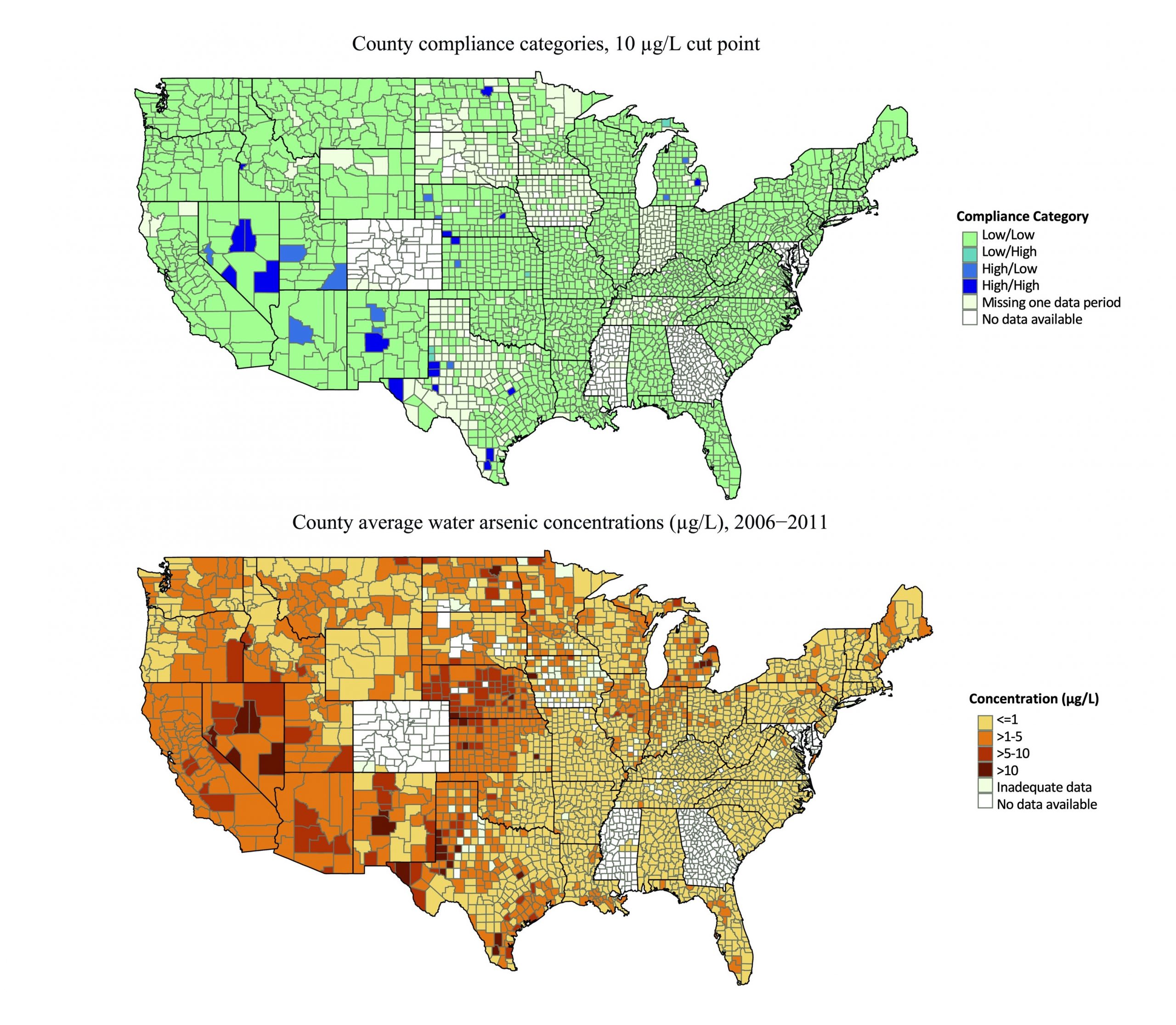
Figure: Maps of counties in compliance with the EPA’s maximum arsenic concentration cut off of 10ug/L (top) and the average water arsenic concentrations across a six year period (bottom). Top Map: Low/Low: less 10μg/L over the six years; High/Low: greater than 10μg/L in 2006–2008, but less than 10μg/L in 2009–2011; Low/High: less than 10μg/L in 2006–2008 but greater than 10μg/L in 2009–2011; and High/High greater than 10μg/L in both periods. Figure was adapted from Figure 3 and Figure 4, Nigra et al., 2020 


Dr. Anne Nigra is a current assistant professor and previous postdoc  in Environmental Health Sciences at Columbia University’s Mailman School of Public Health. 

Reviewed by: Molly Scott, Maaike Schilperoort

Covid-19 and Immunity after Organ Transplant

Over the last two years, the SARS-CoV-2 (Covid-19) pandemic has been at the forefront of media coverage. Hospitals have been overwhelmed, full cities locked down, travel banned, and we are all desperately waiting for a return to the normalcy that immunity promises. However, the development and retention of immunity can be dependent on the individual, and Covid-19 has been particularly daunting to individuals  with weakened immune systems (people who are immunocompromised). These individuals are at an increased risk of succumbing to Covid-19. Overall, it has been easy to identify the individuals that fall into this risk category. However, there has been limited research on the immunity of individuals that  have undergone organ transplants. In a new article by Dr. Mithil Soni, researchers have identified the effects of a solid organ transplant (SOT) on the development and retention of immunity to a plethora of viruses including SARS-CoV-2. SOTs are transplants that  include the kidney, liver, heart, lungs, intestines, and pancreas .

 Dr. Soni and colleagues also focus on the immunity generated by T cells, or cells beyond antibodies that play a role in killing viruses that enter the body. In this study, they focused on the immune response of one patient, a 33-year old male  suffering from erythropoietic protoporphyria, a genetic metabolic disorder that results in excessive liver damage. This subject underwent a SOT and received a liver. When undergoing a SOT, individuals are usually put through a stringent course of immunosuppressants to prevent organ rejection, which places them in the category of immunocompromised.  To their surprise, during a check-up this patient was found to have antibodies for SARS-CoV-2, indicating a previous Covid-19 infection without any serious symptoms. The ability to overcome Covid-19 with minimal symptoms while being classified as immunocompromised intrigued Dr. Soni and colleagues, and the patient agreed to provide his blood for further testing.  

The team went on to test the patient’s immune response to many infections that usually impact immunocompromised individuals. They tested the blood’s immune response to cytomegalovirus and BK virus, two viral infections that immunocompromised and SOT patients are prone to. They also tested the response to Epstein-Barr virus, which can cause Mononucleosis. From the blood, Dr. Soni and colleagues were able to collect and grow the T cells in their lab, exposed them to viruses, and measured their release of cytokines, proteins that are important for a strong immune response. They found a very strong T cell immune response against both cytomegalovirus and BK virus. They also tested the immune response to SARS CoV-2 and other coronaviruses and found a similar level T cell immune response as seen with cytomegalovirus and BK virus. 

These findings overall indicated that the SOT patient continued to have a robust immune response to multiple viruses despite the immunocompromised status. This study shows that it is possible to have robust immune responses to viruses including SARS CoV-2 in an immunocompromised state such as seen after a SOT. However, this research is based on a single case study. To truly understand T-cell memory and activity in immunocompromised individuals much more research has to be done. This means Dr. Soni and colleagues still have their work cut out for them and are actively expanding the research done here. Their next immediate steps are to repeat this study with blood from a larger group of healthy and immunocompromised individuals in the hopes that they will eventually be able to answer the question of SOT and immunity.

Figure: Depiction of increased immunity after SOT.  Top: Liver transplant. Bottom: Expected T cell activity in response to virus vs actual T cell activity in response to virus. 


Dr. Mithil Soni, is a previous Postdoctoral Research Fellow and current Associate Research Scientist at Columbia University.

Take a Break: How the Brain Chooses When to Explore and When to Rest

Have you ever wondered why we feel comfortable in a familiar place or why going back to our favorite spots over and over again feels so good? Well, Dr. Paolo Botta, a former postdoc at Columbia University, and colleagues attempted to unravel some of the inner workings of the brain when it comes to rest and exploration. More specifically, Dr. Botta examined how neuronal activity correlates with periods of rest when exploring new areas. Dr. Botta and colleagues followed the behavior of mice as they freely explored a new area. They specifically looked at where and how often these mice decided to exhibit arrest behavior, or, in other words, take a break during their explorations. While the arrest behavior alone is a fascinating phenomenon and provides insight into how mice explore new spaces, Dr. Botta and colleagues decided to go a step further and see which neurons in the brain are important for this arrest behavior. They decide to home in on an area of the brain called the Nucleus of the Basal Lateral Amygdala (BLA). This area has previously been shown to be involved in locomotor exploration, experience based learning, recognition of familiar areas.

With this information in hand, Dr. Botta and colleagues began by identifying whether BLA neurons are active during arrest behavior. To this end, they gave mice access to both their home cages and a large open area for five days and allowed them to freely explore the large open area during this period. BLA neuronal activity was monitored in the mice by measuring calcium levels, with higher calcium levels indicating neuronal activity (Figure). The researchers observed an increase in calcium in BLA neurons during arrest behavior, which means that BLA neurons are involved in this type of behavior.  However, do these neurons actually cause the arrest behavior? To answer this question, Dr. Botta and colleagues either activated or inhibited the neurons using optogenetics. Optogenetics is a technique in which neurons are stimulated by light. So, by turning different lights on and off, the researchers were able to either activate or inhibit BLA neurons whenever they wanted to. When they activated the BLA neurons, the mice decreased their speed and experienced more arrest behavior. When they inhibited the BLA neurons, the mice had an increase in movement speed. After seeing how turning BLA neurons on and off affected behavior, they concluded that the BLA neurons are important for inducing arrest behavior.

At this point, Dr. Botta and colleagues have revealed that BLA neuronal activity occurs specifically during these arrest behaviors and that their activity is important for the onset of the arrest. However, their curiosity did not stop there. They began to wonder whether BLA activity changed when the mice exhibited arrest behavior, or took breaks in more familiar areas. To figure this out, Dr. Botto and colleagues tracked exactly where the mice explored and counted how many times the mice exhibited arrest behavior in areas that they previously explored. With this experiment, they realized that the mice were more likely to exhibit arrest behavior in areas previously visited. So, mice, like humans, have favorite spots and they like to rest in those spots! After seeing that the mice have favorite spots, Dr. Botto and colleagues went on to examine the BLA neuronal activity in these familiar areas. They found that there was an increase in neuronal activity in these familiar areas and the more a mouse revisited and exhibited arrest behavior in a specific area the more neuronal activity developed. In other words, the more often a mouse took a break in a specific area the more that correlated with BLA neuronal activity.

The amygdala has multiple nuclei, which consist of groups of cells that are important for specific roles. The Central Nucleus of the Amygdala (CEA) is a part of the amygdala that has previously been shown to be involved in immobility. BLA neurons also communicate with the CEA (Figure). Knowing that the BLA neurons are important for invoking arrest behavior and the CEA plays a role in immobility, Dr. Botta and colleagues were curious as to whether these BLA neurons that project to the CEA are the specific neurons involved in triggering arrest behavior. To see whether the BLA neurons that project to the CEA are the ones active during arrest behavior they used the combination of calcium imaging and optogenetic techniques previously mentioned. With these techniques they were able to see that the BLA neurons that project to the CEA had an increase in neuronal activity during arrest behavior (Figure). This increase was not seen in BLA neurons that projected to other parts of the amygdala indicating that the BLA-CEA interaction is integral for the arrest activity. They also repeated the stimulation of the BLA neurons that project to CEA and observed an increase in arrest while inhibiting the same neurons resulted in an increase in movement, further confirming the need of this BLA-CEA interaction to induce arrest behavior.

Overall, Dr. Botto and colleagues discovered that BLA neurons that communicate with the CEA are important for arrest behavior, particularly in familiar places. This behavior seems to be extremely important for allowing a mouse to orient itself and properly explore novel surroundings. Maybe humans have a similar pathway that we use when wandering around. Could my BLA be the reason why I always go to the same cafes after a long walk or stop in the same part of the park while walking my dog? Are our BLA neurons just firing away while we rest? 


Figure: BLA neuronal activity during exploratory vs arrest behavior.      Left: Decreased activity in BLA neurons that communicate with the CEA results in increased exploratory behavior. Right: Increased BLA to CEA neuronal activity, indicated by calcium signaling, results in increased arrest behavior. Red colors indicate decreased BLA neuronal activity and increased exploratory behavior. Green colors indicate increased BLA neuronal activity and increased arrest behavior. BLA: Nucleus of the Basolateral Amygdala, CEA: Central Nucleus of the Amygdala

The Different Perceptions of Cultural Appropriation

The term cultural appropriation, is by far a familiar one. It is defined as situations where a person associated with one group uses cultural elements from another group. These elements can include cultural items like “symbols, genres, expressions, technology and artifacts”. While the term is widely used, actual empirical data surrounding the perception of cultural appropriation is limited. In a recent publication, Dr. Ariel Mosley, a Columbia postdoc, and Dr. Biernat venture into the perception of cultural appropriation. To understand how cultural appropriation is perceived by different groups, Dr. Mosley utilizes an approach of a majority and a minority group in the same community and identifies how each group views different actions as cultural appropriation. 

This study uses multiple examples of cultural appropriation (Figure) to identify the perception of appropriation (whether the example is actually cultural appropriation), perception of harm (whether the appropriation can be harmful to the group the cultural aspect was borrowed from), perception of intent (whether the appropriation was done purposefully), and distinctiveness threat (whether the appropriation threatened cultural aspects that allow the minority group to be distinct from the majority group).

To fully identify the perception of cultural appropriation, this study was divided into five sub-studies. Studies one through three focused on the perception of appropriation, harm and intent, study four focused on manipulating distinctiveness threat, and study five focused on fully crossing the actor and race. They recruited an equal number of adults that either identified as Black or White Americans, with White Americans being considered representative of the majority group and Black Americans representative of the minority group. For studies one through three, the authors set out to answer whether Black Americans or White Americans would have higher perceptions of appropriation, harm, intent, and distinctiveness threat. They used a design where the participants would read scenarios, adopted from social media and news clips, of potential cultural appropriation. In these scenarios the perpetrator, the person doing the appropriating, could be either white or black. The participants were asked to review six possible cases of cultural appropriation (Figure). Throughout the three studies they found that Black participants perceived more cases of appropriation than White participants when the perpetrator was White. In a similar pattern, Black participants saw the scenarios as more harmful, and with intent when the perpetrator was White. When the perpetrator was Black neither White participants nor Black participants saw the scenario as appropriation. Black participants also overall felt an increased distinctiveness threat when compared to White participants. These findings supported Dr. Mosley and Dr. Biernats’ original hypothesis of cultural appropriation being more likely to be perceived when perceivers were members of the minority group.

Since in studies one through three, Black participants felt an increased distinctiveness threat, Dr. Mosley and Dr. Beirnat wanted to see whether increased distinctiveness threat in particular could alter the perception of cultural appropriation. To test this the authors primed the participants in a fourth study for increased distinctiveness threat and focused on one scenario category, “hairstyle” (Figure). They primed the participants to either have increased distinctiveness of threat by having them read, “The Disappearing Color Line in America” or normal distinctiveness of threat by having them read, “The Geography and Climate in America”. Black participants were widely unaffected by the priming with the results mimicking studies one through three, but for White participants, those that were primed for distinctiveness of threat saw the White perpetrators’ actions as cultural appropriation. These results indicated that the level of distinctiveness threat experienced increases the perception of cultural appropriation.

Figure: Detailed depiction of the study designs and categories of cultural appropriation.

Then in study five, to reassure their results, the authors paired a perpetrator with a product that was distinctly part of the participant’s culture. The previous four studies used an item/product that was outside of the perpetrator’s culture, but not necessarily an item belonging to the participant’s culture.  Here they used an item/product that was explicitly part of the participant’s culture. The perpetrator was either a White waiter serving culturally Black cuisines or a Black waiter serving culturally White cuisines. Mimicking their previous studies, they found that Black volunteers were more likely to see cultural appropriation when the waiter was White.

Overall, their study indicated that majority and minority groups perceive cultural appropriation differently, with the minority group being more sensitive to actions that can be perceived as appropriative. They also found that harm and intent correlated with appropriation leading them to the conclusion that both perceptions are part of the appropriation construct. These findings supported their initial hypothesis that power relations and social constructs affect the perception of cultural appropriation and added empirical data to a topic often spoken about but yet understudied.

While Dr. Mosley and Dr. Beirnat have added a significant amount of empirical information on how cultural appropriation is perceived, there is still more to explore. Future studies could expand on how cultural appropriation affects multiple other groups including individuals across different races, sexual orientations, genders and individuals with disabilities. 


Dr. Ashley Mosley is a Post-Doctoral Research Scientist in the Department of Psychology at Columbia University. Her research focuses on social cognition, social identity and intergroup biases. More information about Dr. Mosley can be found on her website.

Follow this blog

Get every new post delivered right to your inbox.