A small investment could reap a big reward

It is well-established that children growing up in economically disadvantaged circumstances can experience a wide variety of challenges to their development. For example, in the domain of language, psychological research shows that even before children reach the age of 3, those from lower socioeconomic (SES) backgrounds hear fewer and less complex words from caregivers compared to their more advantaged peers. This phenomenon places these children at risk of later language learning difficulties. Research groups and other stakeholders around the globe are working tirelessly to craft intervention programs, in a variety of contexts, to target the optimal development of children from under-resourced environments. To target language outcomes, for instance, an initiative called Talk With Me Baby aims to help parents learn about and engage in crucial linguistic interactions with their children from their earliest days.

One prominent intervention in this field is taking a different approach. The Baby’s First Years (BFY) project, directed by a team of leaders in the realms of neuroscience, psychology, and economics, provides families living in poverty with cash and is studying how this money might impact developmental outcomes. These scientists recruited 1,000 mothers from around the US just hours after their babies were born. Half of the mothers are assigned to a high-cash gift group ($333 per month) and the other half are assigned to a low-cash gift group ($20 per month). These payments come in the form of a debit card and the moms are allowed to spend these funds however they’d like.

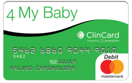
The “4 My Baby” card, the debit card that BFY mothers receive. Courtesy of Baby’s First Years.

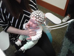
After one year of these payments, the researchers went into the homes of the families and measured a variety of both maternal and child outcomes. One of these measures was EEG, or electroencephalography, to capture the brain activity of the infants. EEG measures the electrical activity occurring between cells in the brain. The findings, published in a recent paper in PNAS by lead author and Columbia postdoc Dr. Sonya Troller-Renfree, captured global attention. She and her team found that infants whose mothers were assigned to the high-cash gift group displayed more high-frequency brain activity compared to children in the low-cash group. Importantly, previous research links this high-frequency activity to the development of thinking and learning. Although the evidence is not air tight, Dr. Troller-Renfree and her team are the first to show there may be a causal link between poverty reduction and changes in brain activity.

There are many pathways through which the gifted money might be impacting children’s brains, which is currently under investigation. It is important to understand that this study is still ongoing and the children in the sample are all currently around 3-years-old. The research team plans to assess the mothers and preschoolers at age 4 on a variety of outcomes. This finding on the potential link between poverty reduction and the brain, along with other work demonstrating that economic support for parents could greatly benefit children’s outcomes, has important implications for public policies that support families and children. While the US has a long way to go in supporting its youth, growing evidence indeed supports the idea that relatively minor investments in children can positively impact their trajectory.

Dr. Sonya V. Troller-Renfree is a Goldberg Postdoctoral Fellow in the Neurocognition, Early Experience and Development Lab at Teachers College, Columbia University. Her research focuses on the effects of early adversity and poverty on cognitive and neural development. She intends to continue examining these questions as part of her new, federally-funded Pathway to Independence Award (K99/00). You can stay up-to-date on her research findings on Twitter at @STRscience or on her website: www.sonyatrollerrenfree.com.


Identifying a potential risk factor for alcohol abuse among victims of violence in childhood

Half of all children in the United States have been physically assaulted in their lifetime, according to a 2014 study. This finding is alarming, especially considering that childhood maltreatment and abuse can lead to numerous negative mental health outcomes. 

Researchers and medical professionals around the globe often focus on adverse childhood experiences and their detrimental effects on development. Experiencing threat and violence is frequently correlated with a decreased ability to effectively handle negative emotions and heightened emotional reactivity relative to those who have not experienced such trauma. For instance, a typical situation such as having your toy taken away by a peer in school might invoke an explosive, angry response from a child who has been a victim of abuse. Moreover, research demonstrates that children who have faced abuse are also more likely than others to interpret ambiguous actions (such as a classmate accidentally bumping into them in the hallway) as confrontational.

How might those who’ve experienced violence in their childhood, and also have trouble dealing with negative emotions, respond to everyday stressors (i.e. getting through hard homework sets or dealing with long waits for customer service on the phone)? Dr. Charlotte Heleniak and her colleagues studied this response, called distress tolerance, in a newly published paper.

Levels of distress tolerance vary among different individuals. Someone with low distress tolerance is extraordinarily uncomfortable in situations where they’re facing a challenging obstacle, upset, or experiencing negative emotions that can make it hard to persist in the face of difficulty. They have a harder time working through these difficult events compared to people with higher distress tolerance. Research also shows that people with low distress tolerance may find it necessary to escape bad feelings by seeking immediate relief. This relief can often take the form of substance abuse. 

Additionally, while little research has been done, distress tolerance may make an individual more vulnerable to other mental health problems such anxiety and depression. Because of this, Dr. Heleniak’s team examined whether low distress tolerance is associated with these two mental illnesses, as well as alcohol abuse.

teens drinking
Image from Pixabay

Propensity toward problematic alcohol use in adolescents involves many environmental risk factors such as sociodemographic factors and parental drinking behavior. These can be difficult or impossible to address therapeutically. However, if distress tolerance is indeed tied to substance abuse, this may offer a clearer path toward crafting a psychological intervention. 

Dr. Heleniak and her colleagues studied 287 16- to 17-year-old participants across a broad range of socioeconomic backgrounds. They asked the teens about previous violence exposure in their personal life, and assessed depression, anxiety, and alcohol use. Four months later, they reassessed these parameters.

To examine the teens’ distress tolerance, the researchers used a measure called the Paced Auditory Serial Addition Task, which measures a person’s persistence on a difficult task. The sooner a participant decides to terminate the task, the lower their distress tolerance. The team found that those teens who experienced a heighted amount of violence did indeed have lower distress tolerance. At the initial time point, lower levels of distress tolerance were not associated with any of the three psychopathologies (i.e. alcohol abuse, anxiety, depression).

However, the researchers found that low distress tolerance predicted alcohol abuse from the first time point to the second, about 4 months later. Low distress tolerance was not associated with anxiety or depression at either of the two time points of data collection.

Figure 1. Teens who experienced more abuse and violence had lower distress tolerance. Four months after the initial assessment, teens who had low distress tolerance were even more likely to have developed problematic drinking behaviors.

Based on their findings, Dr. Heleniak and her team conclude that researchers could potentially pinpoint distress tolerance as a way to target teens’ problematic use of alcohol, especially those who have experienced violence. Indeed, therapeutic programs aimed at improving distress tolerance already exist. The authors explain that treatments such as Dialectical Behavior Therapy (DBT) and mindfulness practices may be particularly useful. 

Given that teen alcohol abuse may continue into adulthood and lead to dependency issues later in life, the findings of this study could go a long way to helping those adolescents who struggle with both addiction issues and an abusive past.

If you or someone you know is experiencing substance dependency problems, SAMHSA (1-800-662-HELP) is a free, confidential, resource available 24/7 365-days a year.

Charlotte Heleniak is a postdoctoral scientist in the Developmental Affective Neuroscience Lab at Columbia University. She received her Ph.D. in Child Clinical Psychology from the University of Washington. She focuses on how childhood adversity impacts emotion regulation and social cognition in ways that predict adolescent psychopathology. This research has earned her awards from the National Institute of Mental Health and the Doris Duke Charitable Foundation, as well as the Sparks Early Career Grant from the American Psychological Foundation.

A pain in the foot: moves to prevent injury in dancers

Dancing can be one of life’s greatest pleasures. But for folks who consistently engage in intensive forms of dance, such as ballet, it can also lead to injury. One injury amongst dancers and other athletes is flexor hallucis longus tendinopathy

The flexor hallucis longus tendon (FHL), as seen in Figure 1, helps stabilize a person when they’re on their toes and mainly moves to flex the big toe. It stretches all the way from the calf muscle, through the ankle, down to the big toe. When athletes engage in repetitive movements that recruit their foot and ankle in this manner, like jumping up and pushing off the big toe, strain of the FHL tendon can occur. FHL tendinopathy is painful and can leave dancers and gymnasts out of commission from their passion and profession.

The posterior view of the FHL in the right leg, taken from Sports Injury Bulletin
Figure 1. The posterior view of the FHL in the right leg, taken from Sports Injury Bulletin.

Luckily, researchers study overuse conditions like this one. Dr. Hai-Jung Steffi Shih and her colleagues recently published a study where they had 17 female dancers (9 with FHL tendinopathy and 8 without) perform a specific ballet move called saut du chat. The dancers wore a full-body marker set that allowed the researchers to capture the fine-grained positions and movements of the dancers’ bodies throughout the ballet move. 

When performing a movement like the saut du chat, the body tends to place a heavy load on one particular joint in the foot called the metatarsophalangeal joint (MTP). Repeating the movement like this over and over again (as dancers often do), can contribute to overuse of the FHL. Researchers can measure something called stiffness in an athlete’s musculoskeletal system to assess the potential for injury. Scientists think that greater stiffness may lead to impact on a person’s bone, while reduced stiffness may lead to soft tissue injury. To better understand stiffness, it can help to think about parts of the lower body as springs, such as the ankle, knee, and hip joints. They compress, store energy, and then release – like when you squat and jump. Researchers can examine stiffness of these joints, specifically joint torsional stiffness, or how easy or hard the joints bend.

Additionally, researchers who study movement can also measure how a dancer’s body makes contact with the ground to determine if certain kinetic factors might be significantly associated with injury. For instance, the difference in the angle at which a dancer’s lower limb makes contact with the ground has been associated with injured and uninjured groups of dancers. If researchers can accurately indicate which angles are associated with injury, they can collaborate with medical professionals and teachers to craft targeted interventions on how a dancer should be properly moving their body.

In the current study, the researchers posited that lower extremity joint torsional stiffness measured when participants made contact with the ground would be altered in dancers with tendinopathy compared to uninjured dancers. They also thought that dancers with tendinopathy would demonstrate a lower limb posture associated with kinetic factors that differed between injured and uninjured dancers. 

Using the marker set that the dancers wore, the researchers measured torsional stiffness by examining the rotational forces, or the change of joint movements, over the change of the joint angle over a period of time. This data was gathered as the dancers flexed and extended their lower extremity joints during the saut du chat. Moreover, the team measured the contact angle at which the dancers’ feet took off from. 

Dr. Shih and her colleagues found that the dancers with tendinopathy demonstrated less joint torsional stiffness at the metatarsophalangeal (MTP), ankle, and knee joints during the takeoff step of the dance move. To reiterate, research suggests that a lack of joint stiffness is not good because it could lead to soft tissue injury, as it allows for excessive joint motion. Additionally, these injured dancers took longer to reach peak force when pushing off the ground and the peak force was also lower than in the uninjured dancers. Finally, the angle at which dancers first contacted the ground during that take off step was smaller (i.e., their foot was further in front of their pelvis) in participants with FHL tendinopathy compared to those without injury.

How can these findings help dancers and those that provide movement guidance to dancers? Knowing the particular biomechanical changes that precede tendinopathy can inform targeted interventions aimed at improving how dancers move their feet and legs when performing certain moves. For example, teachers can offer cues and guidance on how a dancer should position their pelvis and feet in an effort to prevent injury. 

This study by Dr. Shih and her colleagues is the first to demonstrate the differences in movement profiles between dancers with and without FHL tendinopathy and could go a long way to informing interventions that could prolong dancers’ careers.

Dr. Hai-Jung Steffi Shih is currently a postdoctoral research fellow in the Neurorehabilitation Research Lab at Teachers College, Columbia University. She received her PhD in Biokinesiology and Physical Therapy at the University of Southern California where this research was conducted. Steffi’s research aims are to further understand musculoskeletal pain, movement disorders, and improve intervention strategies using a multidisciplinary approach. Outside of academia, Steffi is an avid traveler who has been to more than 35 countries. She loves to dance, enjoys playing music, and is aspiring to become an excellent dog owner one day. You can email her at [email protected] and connect with her on Twitter @HiSteffiPT.

Meet Olaya Fernandez Gayol; Postdoc in the Department of Pediatrics

We’re back with our ‘Meet the Postdocs’ Series! Today we’re featuring our fearless and incredible CUPS co-President, Olaya Fernandez Gayol.

Olaya Fernandez Gayol, Postdoctoral Research Scientist in the Department of Pediatrics

How do you identify? What should we call you?

I identify as a queer woman (pronouns she/her/ella) and my name is pronounced oh-LAH-yah.

Which department are you in at Columbia and what is your position?

I´m a Postdoctoral Research Scientist in the Department of Pediatrics (Division of Molecular Genetics). I’m located in the 6th floor of the Russ Berrie Medical Pavilion in the CUIMC campus.

Where are you from and how long have you been in NYC? 

I’m from Spain (from a city in the north called Oviedo) and I have now been in NYC for a little over 2.5 years.

Where did you go to school? Describe your path to your current position.     

I did my undergrad in Biology, Master and PhD in Neuroscience all at Universitat Autònoma de Barcelona (Spain). During my PhD, I got a scholarship to come to the US for 3 months to the lab of Dr. Richard Palmiter (University of Washington – Seattle). My PI knew him from previous collaborations, so it was pretty easy to set up. While I was there creating a new transgenic mouse (read about it on this post), Dr. Stephanie Padilla (then a postdoc in the lab) took me under her wing and we worked together on a little project on Agrp neuron activation. Four years later, she reached out to me to tell me that her former PhD advisor, Dr. Lori Zeltser, was looking for a postdoc and that she had recommended me. I interviewed with Lori a couple of times and got the job! I found the project on brain circuits related to anorexia nervosa very exciting and a good change after doing a PhD mostly on obesity. Also, New York City.

What research question are you trying to figure out right now?

I’m working on a couple different projects. My main project is about understanding the role of neurons that produce brain-derived neurotrophic factor (BDNF) in a small area of the brain called the amygdala, and how a very common mutation in humans affects their activity, food intake and stress. The second question I’m tackling is to determine how another mutation (this one in vasopressin receptor 1A), which has been identified in human patients with anorexia nervosa, impacts its activity.

In a nutshell, what tools or approaches are you using to try and figure this out?

I mostly use molecular biology techniques like reverse transcription real-time PCR (DNA amplification from RNA), Western blot (protein quantification) and immunofluorescence (staining of tissue samples) using tissue from mice (both genetically modified and wild type). Recently, I started using cell culture approaches with human cell lines to study the activity of the vasopressin receptor.

What is the best part of your job?

Being so close to first-class researchers who can help me learn new and exciting techniques and being able to attend their talks as well.

Why do you love science?

I really enjoy the troubleshooting part of science, the thrill of getting a new protocol to work to answer your questions!

What advice would you give to people interested in a career in science?

If you are fascinated by discovering new things, science can be a great outlet for that. However, be aware that there is science beyond academic research, this is only a tiny part of the scientific world, and you really need to be aware of what it entails (TALK to people!). Explore other possibilities, there is so much you can do “in science” and not all of it needs to fit the image of the detached from reality scientist we’ve been exposed to.

Tell us a bit about yourself or your projects that are not related to science. 

I love musicals, cooking and baking (it’s like science!), hiking and playing board games. A lot of my free time is devoted to either those, or working with CUPSECUSA (a Spanish association for scientists in the US) and the NPA.

What is your favorite thing about NYC?

Broadway, duh (September 2021 cannot come soon enough). Apart from that, I really enjoy visiting museums and having picnics in the awesome parks. I also appreciate that it’s easy to catch a train and go hiking to escape the concrete jungle.

What do you like the most about CUPS? 

The passionate postdocs who make it up! It’s been an exceptional opportunity to meet people I wouldn’t have interacted with otherwise. In addition, it’s a great chance to build leadership and organizational skills. It allows a hands-on approach to your career development, rather than passively attending events you have a say in determining the programing.

How can folks follow you on social media / contact you?

You can engage with me on the CUPS Slack, by emailTwitter or LinkedIn (all of the above is also a valid answer).

Thank you so much, Olaya!

Maternal Stress and the Developing Brain

As humans, we all experience stress. It is a normal, and sometimes even beneficial, part of life. A small amount of stress can help motivate someone to prepare for a job interview or study for an important exam. There are times, however, when stressors become too overwhelming and even detrimental to health. Scientists, from medical researchers to psychologists, have studied stress for decades and documented some of these negative impacts on the brain. When thinking about the importance of the foundational, early years of a person, the presence or lack of stress can play a crucial role in development. For instance, extensive research shows that living in poverty is extraordinarily stressful for families and can negatively influence children’s brain development. The impacts of stress resulting from situations such as growing up in poverty warrant further investigation, especially considering that in 2020, one in six children in the U.S. was living in poverty.

Researchers can use various methods to assess how factors like stress impact the brain of growing children. Developmental scientists can use a tool called EEG, short for electroencephalography, to study the brain. EEG measures electrical activity in the brain by recording the communication between brain cells. It is an ideal neuroimaging method for understanding infant brain development since it allows for infants to be awake and moving, and even sitting on their caregiver’s lap during recording. Besides being infant-friendly, EEG is a useful tool for looking at brain development, given that there is a known pattern of how brain activity changes across the first few years of life.

Specifically, when using EEG to look at brain development, scientists typically see two different patterns. Broadly, infants have a mix of different types of brain activity that we call low-frequency and high-frequency power. Low-frequency power (e.g., theta) tends to be higher when the brain is at rest, while high-frequency power (e.g., alpha, beta, and gamma) tends to be used for more complex thinking like reasoning or language. As infants grow, scientists see that low-frequency power decreases and high-frequency power increases. Importantly, we can use EEG to assess how factors like stress impact the tradeoff of low-frequency and high-frequency power in the developing brain.

Image of a one-month-old infant with an EEG cap.
Figure 1. A one-month-old infant with an EEG cap. Courtesy of the Neurocognition, Early Experience and Development Lab.

Research shows that children growing up in chronically stressful environments often show alterations in the typical pattern of brain activity development. To further understand the mechanisms underlying this pattern of development, scientists have begun to study which biological and environmental factors may be at play. For instance, researchers can examine the role of caregiver stress, socioeconomic status, home environment, and neighborhood factors, just to name a few.

A recent paper by Dr. Sonya V. Troller-Renfree and colleagues examined maternal stress by looking at the amount of stress hormone (cortisol) found in hair. This measure assesses chronic stress and provides researchers with the average cortisol level of the mother from the preceding 3 months. Dr. Troller-Renfree’s research group hypothesized that infants who have mothers with higher stress hormone, compared to mothers with lower levels of stress, would show differences in their brain activity. Specifically, the researchers predicted that infants of more chronically stressed mothers would exhibit proportionally more low-frequency power and proportionally less high-frequency power compared to infants with physiologically less-stressed mothers.

Indeed, their results showed that infants of mothers who had higher levels of hair cortisol demonstrated higher levels of low-frequency (theta) activity and lower levels of high-frequency (alpha and gamma) brain activity. This finding is consistent with previous research showing that stress and adversity impacts early neural development. Importantly, Dr. Troller-Renfree’s team sampled a diverse pool of participants (both in terms of socioeconomic status and race), therefore bolstering the generalizability of their findings.

So what are the implications of these alterations? Research suggests that similar patterns of neural activity are associated with negative outcomes later in a child’s life, including language development and psychiatric problems. Nevertheless, this does not mean that a child will undoubtedly experience these issues. Additionally, it may be possible that these patterns, while associated with negative outcomes in some areas, may also be adaptive in other circumstances. Furthermore, the issue of the mechanisms by which a mother’s stress impacts the developing child still remains unclear. How exactly does a mother’s stress level impact the brain of her child?

Based on previous research by other scientists, Dr. Troller-Renfree posits a few mechanisms that must be further explored. For example, it is possible that stress impacts crucial mother-child interactions. It could be that stress hormones are passed from mother to baby in utero or through breastmilk. Moreover, it is also possible that environmental factors impact stress and brain development.

It is crucial that developmental scientists continue studying these mechanisms so that targeted intervention programs can be formed for families facing stress. Indeed, the esteemed pediatrician and researcher Dr. Jack Shonkoff of the Center on the Developing Child said in an episode of The Brain Architects Podcast: “In fact, one of the cardinal principles of the science of early childhood development is that if we want to create the best kind of environment for learning and healthy development for young children, we have to make sure that the adults who care for them are having their needs met as well.” As a society, we must recognize how detrimental stress can be to the developing child and invest in finding effective ways to alleviate caregiver stress.

Dr. Sonya V. Troller-Renfree is a Goldberg Postdoctoral Fellow in the Neurocognition, Early Experience and Development Lab at Teachers College, Columbia University. Her research focuses on the effects of early adversity and poverty on cognitive and neural development. She intends to continue examining these questions as part of her new, federally-funded Pathway to Independence Award (K99/00). You can stay up-to-date on her research findings on Twitter at @STRscience or on her website: www.sonyatrollerrenfree.com.

Follow this blog

Get every new post delivered right to your inbox.