Cuttlefish, octopus, and squid are fascinating marine creatures that have perfected the art of camouflage underwater. With uncanny speed, these soft-bodied cephalopods can change the color, pattern, and texture of their skin to blend seamlessly into their surroundings, impressively adapting to their environment. Unlike many animals, cuttlefish don’t rely on fur or feathers to hide in the background. Instead, they actively manipulate thousands of pigment cells in their skin to acquire the color of the environment around them. This intricate disguise process starts in their brains, as camouflage is a response to the animal’s perception of the external world. To conceal their bodies, cephalopods convert visual inputs into neural representations within their brain, ultimately transmitting signals all the way to the skin, where thousands of tiny structures called chromatophores adjust to allow color changes. However, camouflage is just one captivating aspect of cuttlefish biology. These marine animals present a rich repertoire of signaling behaviors for mating and communication and they are proficient learners, with memory capabilities not often seen in invertebrates. Collectively, these attributes position cephalopods as extraordinarily captivating subjects within the realm of biological studies.
Researchers at Columbia University’s Zuckerman Institute, led by Dr. Richard Axel, have made significant progress in comprehending the shape-shifting abilities of cuttlefish. In a recent article published in Current Biology, they generated a detailed neuroanatomical brain map, revealing insights into how their skin transformation is controlled. Tessa Montague, PhD and colleagues focused on the dwarf cuttlefish (Sepia bandensis), a small tropical species found around coral reefs in the Indo-Pacific Ocean (Figure 1A). The dwarf cuttlefish is an intriguing model for neuroscience research with its rich array of dynamic social behaviors and camouflage. Through an advanced imaging technique called MRI (magnetic resonance imaging), computer programming and web design they constructed a 3D atlas illustrating the dwarf cuttlefish’s brain anatomy (Figure 1B).
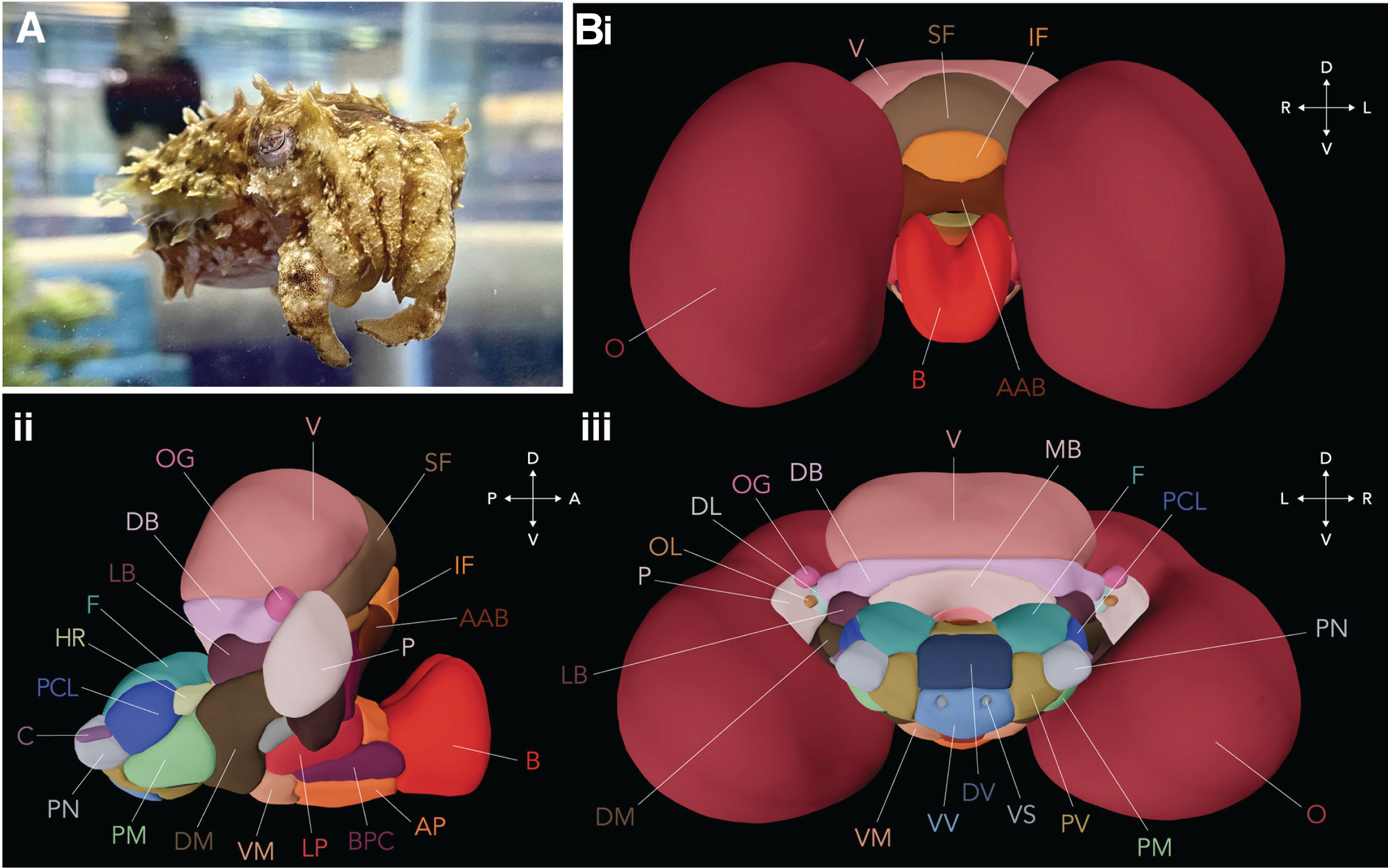 Figure 1. (A) Adult dwarf cuttlefish. (B) 3D template brain, based on magnetic resonance imaging (MRI) of 8 cuttlefish brains, (i) anterior view, (ii) right view, and (iii) posterior view. The abbreviations in color indicate the different brain structures that were identified and mapped. Modified from T. G. Montague et al., 2023
By scanning the bodies and brains of male and female cuttlefish, the researchers identified 32 distinct lobes or functional units within the cuttlefish brain (Figure 1B). Each lobe is densely packed with neurons and performs specialized tasks. The two largest lobes, making up 75% of the total brain volume, are the optic lobes (O in Figure 1B). They receive direct projections from the eyes and process visual information, a crucial step in enabling cuttlefish camouflage. Notably, other key lobes in the camouflage pathway include those controlling the chromatophores, the pigment-filled saccules in cuttlefish skin that provide the color. When the lobes send signals to the chromatophores, these rapidly expand or contract to alter skin shades on a millisecond timescale. The lateral basal lobe (LB in Figure 1B) for example, is the lobe involved in establishing the most appropriate skin pattern components for camouflage. Another brain area highlighted by the atlas is the vertical lobe complex (V and surrounding lobes in Figure 1B), which previous studies suggest plays a key role in learning and memory. Unlocking the functions of this lobe could reveal the neural basis for complex behaviors like camouflage.
While past studies have mapped the brains of related cephalopods like squid and octopus, this is the first complete atlas for a cuttlefish species and it provides the neuroscience field with a valuable comparative perspective. The researchers found strong similarities in the anatomy of the dwarf cuttlefish with the common cuttlefish, despite differences in size and camouflage strategies between the species. This suggests that fundamental aspects of brain organization are conserved, at least among close cephalopod relatives. It also highlights how flexible cuttlefish brains are: they can generate very different camouflage patterns using essentially the same basic circuit layout. Exploring what accounts for such flexibility on the microscopic scale will be the next challenge.
To maximize the utility of the brain atlas, for both educational and research purposes, the authors built an interactive freely available web tool, called Cuttlebase, where users can identify specific brain regions in the histological 2D atlas (that shows the cellular components in the cuttlefish brain) and use the 3D model of the brain to explore the different lobes (Figure 2).
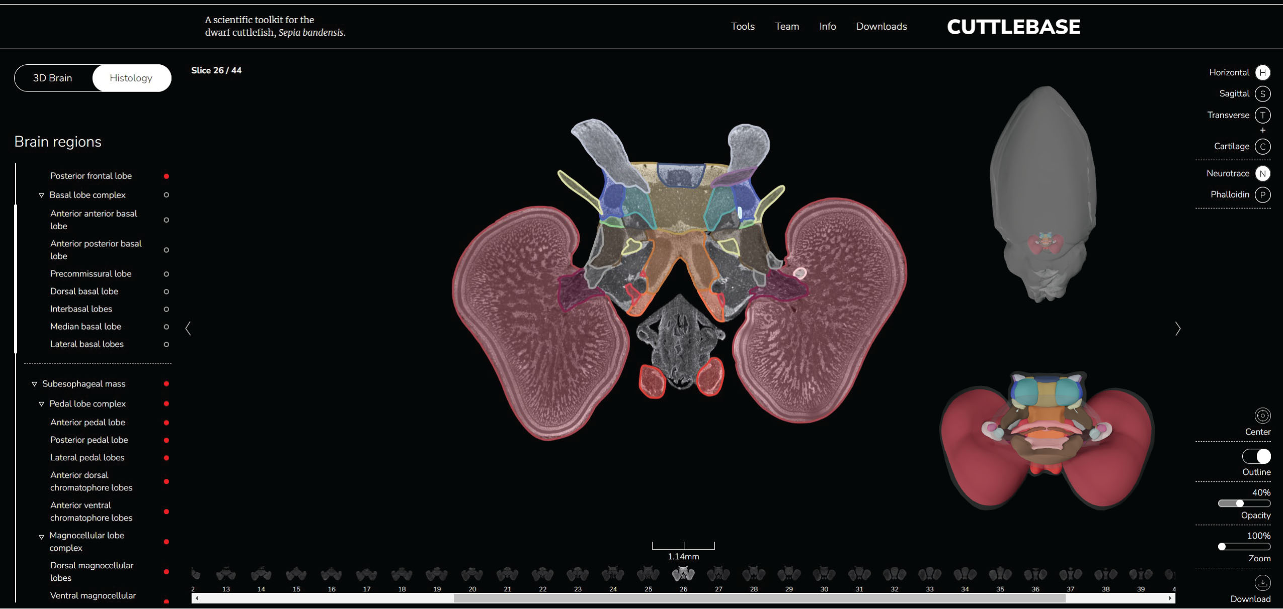 Figure 2. Cuttlebase is an interactive scientific web toolkit for the dwarf cuttlefish that includes a histological brain atlas in 2D and a 3D body and brain atlas to visualize and explore the different organs and brain lobes. Obtained and modified from https://www.cuttlebase.org/
This atlas serves as an invaluable tool for the scientific community to explore the basic anatomical components of complex behaviors and can give us insight into how brains are capable of representing information. It also offers an invaluable anatomy lesson, giving scientists a privileged peek inside the ingenious brains of these undersea masters of disguise. With a detailed understanding of their sophisticated neural systems, unraveling cuttlefish mysteries seems closer than ever.
Reviewed by: Trang Nguyen, Maaike Schilperoort