Newborn octopus neurons steadily march towards maturity from around the eyes into the brain

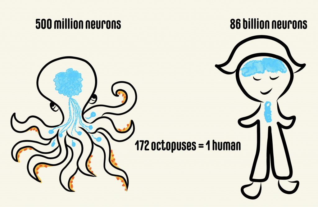
Anyone who watched the movie Arrival would not miss the conspicuous resemblance of the alien Heptapods to some of our own earthly beings – the octopuses. While serving as inspiration for alien creatures in movies, as clairvoyants in soccer World Cup or as savages in classic science fiction and mythology, cephalopods like octopuses and squids have been dubbed as one of the most intelligent creatures on the planet. There is a good reason for why cephalopods, particularly octopuses, have developed such a reputation. Octopuses have a striking organization of brain structure, different from that of any other studied organism. They have the largest nervous system among animals lacking a backbone, comprising a total of nine “brains”. Out of these, one is a major donut-shaped brain that contains ~200 million neurons surrounding the octopus’s food pipe, which is strangely located in the head! This brain communicates behavioral intricacies to the eight so-called mini-brains located within the arms, each containing ~40 million neurons. The central brain is responsible for executing complex behaviors like tool use, ability to plan for the future, shape-shift, camouflage, recognize individuals and solve complex puzzles. While the last common ancestor between octopuses and humans was about 680 million years ago, a recent surprising discovery showed that they both evolved to use the same molecules during development. Scientists discovered that genes that produced the camera-like eye in humans are the same ones that gave rise to the camera-like eye in octopuses. What’s more, these cephalopods have evolved complex brains that show behavioral innovation on par with a small primate.

Comparison between the number of neurons present in the octopus and human brains. The octopus has one major brain and eight “mini-brains” while humans have neurons in the head and the spinal cord.

Despite the potential that the octopus provides for understanding developmental biology, particularly the brain, the molecules that dictate how the mollusk’s brain is built are unknown. The common octopus, Octopus vulgaris, is specifically suited to address this question because it can produce thousands of small and transparent eggs in a single batch and scientists have recently mapped out most of its genes. Using O. vulgaris in studies led by Dr. Astrid Deryckere from Dr. Eve Seuntjens’s lab at KU Leuven in Belgium, the group set out to unravel these molecular mysteries. “If you would think of cephalopods as the primates of the sea, that have evolved a complex nervous system from a far more simple ancestral nervous system, surprisingly little is understood on the morphological and molecular mechanisms driving its development.”, said Dr. Deryckere. They approached the problem in two studies. In the first study, she established a system for controlled embryonic development, which enabled her to care for thousands of eggs without the need for the mother octopus. She used state of the art microscopy and recorded high-resolution images of octopus development from fertilization through hatching. This work from Dr. Deryckere and colleagues can now be used as an elaborate reference for cephalopod embryology. 

O. vulgaris hatchling imaged in 3D at high resolution after labelling DNA (cyan). The same embryo was imaged from different orientations: back view (left), side view (middle) and front view (right). The head is located on top and the arms are at the bottom. Images were produced by Dr. Astrid Deryckere in the lab of Dr. Eve Seuntjens, KU Leuven, Belgium.

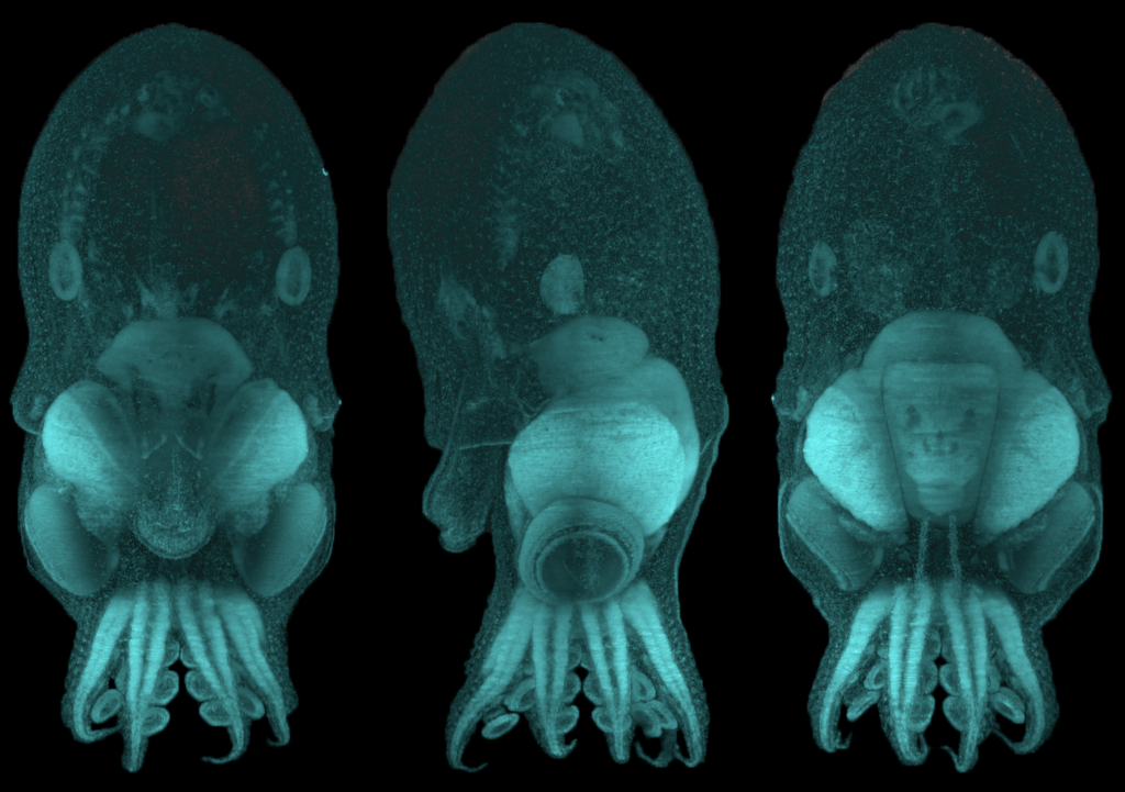
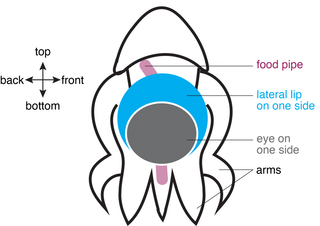
In the second study, Dr. Deryckere dug deep into the origins of the octopus brain. She used precise staging to track the precursor cells of neurons, or “neuronal progenitors”, that generate specialized neurons. Intriguingly, these progenitor cells appeared in structures called the lateral lips, that are unrelated to the brain and are located around the eyes. So, it appeared that neurons were first born in these structures and eventually migrated into the central brain – a possibility that prompted the authors to investigate further. They found that hundreds of thousands of neurons were created within octopus embryos even before hatching. To find out what genes are required for this unique way of making neurons, Dr. Deryckere used molecular markers and showed for the first time that newborn neurons travel long-distances to reach their final location in the brain. The results showed that the genes were expressed in the same order that vertebrates like humans do. By closely observing entire embryos in three dimensions during their growth, she found that neurons proceed through maturation while migrating from the lateral lips to an intermediate transition zone that finally leads to the brain. 

Schematic of O. vulgaris embryo, indicating the location of the lateral lip in relation to the food pipe and the eye. Schematic adapted from images in Deryckere et al., 2021.

Using detailed molecular studies, the scientists now show support for the lateral lips harboring newly dividing neuronal cells in the embryo. This is unusual because unlike in human brains and many other organisms, the dividing cells are located outside the central brain. These dividing cells then unwaveringly make their way towards the final destination to maturity in the octopus central brain. “The migration is especially exceptional for invertebrates where neurons usually migrate only a few cell lengths”, noted Dr. Deryckere about the rarity of the observation.

This unique development in the octopus head and its interesting age-dependent arrangement of dividing cells and mature neurons only inspires further reverence for the cephalopod. While it continues to influence characters in pop culture, the glorious octopus and its brain hold even more promise in the real world. The octopus brain’s cognitive ability has galvanized a new age of artificial intelligence, leading to the construction of flexible robotics and prosthetics, but at the same time, is pushing scientists and philosophers to tackle the important question of how an intelligent life form is defined.

Dr. Astrid Deryckere is currently a postdoc in the lab of Maria Tosches in the Department of Biological Sciences at Columbia University. Her focus remains on brain development but she has transitioned to working on an animal with a backbone – the salamander.

Laboratory evolution of a cellular reprogrammer provides a potent path to stem cell generation

The human body has approximately 15 trillion cells, all of which arise from embryonic stem cells which are considered the building blocks of life. Stem cells renew themselves by dividing indefinitely and can also give rise to cells with specialized functions which ultimately end up forming various organs and tissues in our body. This process is called differentiation. Typically, once cells specialize or differentiate, they lose the ability within the body to go back to being stem cells. Given their unique properties, stem cells have become a critical starting point that scientists can tinker with to develop new drugs and therapies. Because of their tremendous value for research, scientists have figured out non-invasive ways to transform differentiated cells into cells with stem cell like properties. These lab-grown cells, called induced pluripotent stem cells or iPSCs, are typically generated by a process called “cellular reprogramming”.

As Dr. Tania Thimraj explains in a recent article, proteins called transcription factors can act as cellular “fixer-uppers” and renovate differentiated cells to look and behave like stem cells. The current state of the art process for making iPSCs involves excess production, also known as overexpression, of the following transcription factors in differentiated cells: Oct4, Sox2, Klf4, and c-Myc (collectively called the “OKSM” cocktail). Despite significant advances in the formulation of this cocktail, there is still a huge margin for improvement in the ability of this cocktail to transform differentiated cells into stem cells. In a recent study performed by Dr. Tan and co-authored by Dr. Malik, the authors propose that the cocktail is not especially effective because the transcription factors were never under any evolutionary selection pressure to produce stem cells. Inspired by this, the authors set out to use evolution in the dish, also known as directed evolution, to make a more efficacious transcription factor cocktail.

Although natural evolution takes place over millions of years, smaller scale evolution can be done in a laboratory setting at much faster timescales. This approach is known as “directed evolution” and has been successfully used by scientists to evolve proteins with new functionalities. This process involves making random mutations in the protein of interest. Then, these mutants undergo a selection process in an appropriate cellular context so that protein variants with desirable properties can be isolated.

In a pioneering study, members of the Jauch lab, including Dr. Malik, used directed evolution to optimize the cellular reprogramming ability of the transcription factor Sox2. Building on this success, the Jauch lab used directed evolution to make ePOU, an enhanced and evolved version of Oct4 which is an integral part of the OKSM cocktail. In the current study for creating ePOU, the authors made random mutations at six functionally important positions in Oct4 and overexpressed the mutant proteins in mammalian cells such that the Oct4 transcription factor activity was tied to the production of a green fluorescent protein representing stem cell transformation.

This innovative study demonstrates that the transformation potential of naturally occurring transcription factors can be drastically enhanced by directed evolution. In addition, this work also provides a framework for future research on transcription factor engineering for cell reprogramming. By providing a faster and more efficient way to produce stem cells, this study has the potential to accelerate various research and therapeutic avenues such as regenerative medicine, drug efficacy and safety testing, and studying human development and disease.

Dr. Vikas Malik is a Postdoctoral Research Fellow in Dr. Jianlong Wang’s lab in the Department of Medicine at Columbia University Medical Center and is a member of CUPS and the Outreach and Communications Committee.

Transcription factors and cellular fixer-uppers

Self-renewing stem cells are capable of developing into certain specialized cell types thus making them ideal candidates to study human development and as potential treatment modalities for a range of diseases. There are three types of stem cells: embryonic stem cells, adult stem cells and induced pluripotent stem cells. As the name suggests, embryonic stem cells are found in the embryo at very early stages of development. Adult stem cells are found in specific tissues post development. However, using human embryonic stem cells in research is quite restricted due to ethical, religious, and political reasons. This limitation has resulted in the identification of cell reprogramming techniques to convert differentiated cells, such as skin cells, back to an embryonic stem cell state through a process called induced pluripotency. The resulting induced pluripotent stem cells (iPSCs) are equivalent to the natural human embryonic stem cells and can be differentiated to any desired cell type using a mixture of biological molecules.

Cell reprogramming techniques can be likened to fixer-uppers. Imagine trying to remodel a building for a different purpose – converting an office building into a residential one for instance. Though the building material can be reused, with the aid of experts, there would be some structural changes and remodeling necessary to make it a home. Similarly, cellular reprogramming is the technique by which one cell type can be converted to another cell type in the lab with the help of certain gene expression regulators called transcription factors (Fig. 1). The process of inducing pluripotency has been studied extensively and the overexpression of four transcription factors – OCT4, SOX2, KLF4, cMYC (collectively referred to as “OSKM”) – has been shown to induce pluripotency in mouse skin cells.

Many studies have tried to identify other transcription factors with the potential to induce pluripotency or to replace OSKM in an effort to enhance the efficiency of iPSC generation. Of these four transcription factors, SOX2, KLF4 and cMYC have been successfully replaced by members of their protein family to induce pluripotency. However, replacing OCT4 with structurally similar and evolutionarily related factors failed to show similar reprogramming capabilities. This could indicate the presence of special molecular features on OCT4 that give it the ability to reprogram cells. However, these special features and the molecular mechanisms that enable OCT4 to induce pluripotency remain to be identified.

Fig.1. Depiction of pluripotency induction in differentiated cells. Transcription factors regulate the process of converting a mature cell into an induced pluripotent stem cell which can then be directed to differentiate into any desired cell type. Illustration created with BioRender.com

In the current study, Dr. Malik and colleagues hypothesized that the ability of a transcription factor to reconfigure chromatin (the complex of macromolecules composed of DNA, RNA, and protein, which is found inside the nucleus of eukaryotic cells), is one of the features that distinguishes a reprogramming competent transcription factor from a non-competent one (Fig. 2). To test this hypothesis, they studied the well-established OCT4-SOX2 relationship from initiation to maintenance of pluripotency. They performed their study by comparing DNA-accessibility, DNA-binding,  and transcriptional control by OCT4, OCT6 and an OCT4 mutant that does not interact with SOX2 (OCT4defSOX2) during early, mid and late phases of cell reprogramming. What makes this study particularly interesting is the fact that a previous study by the same group has shown that OCT4 naturally interacts with SOX2 to induce pluripotency, whereas OCT6 can only induce pluripotency when OCT6 was mutated to enhance its interaction with SOX2. Dr. Malik’s current study focuses on the mechanisms by which the above-mentioned transcription factors interact with chromatin and in turn bind to the transcription factor binding sites on the genes that are involved in processes from the initiation to maintenance of induced pluripotency.

Fig. 2. Depiction of chromatin remodeling by competent vs non-competent transcription factors. Opening up the chromatin by competent transcription factors and making transcription factor binding sites accessible is required to induce pluripotency. Failure to do so by non-competent transcription factors results in a failure to induce pluripotency. Illustration created with BioRender.com.

From this study, the researchers found that OCT4, OCT6 and OCT4defSOX2 have unique transcription factor binding sites on the pluripotency-related genes which could explain why substituting OCT4 with related transcription factors does not activate these genes. The results from this study challenge previously established roles for OCT4 in driving pluripotency. Dr. Malik and colleagues have identified distinct modes of chromatin interaction and roles for SOX2 and OCT4 during initiation, progression and maintenance of pluripotency. They found SOX2 to be a better facilitator of chromatin opening and initiator of pluripotency compared to OCT4. Once the cells have been initiated towards pluripotency, OCT4-SOX2 binding is required to see the process through and once the cells are pluripotent OCT4-SOX2 binding becomes less essential. The most important role of OCT4, they found, was to maintain the cells in a pluripotent state as opposed to its previously investigated role as an initiator of pluripotency. 

The results from this study contribute new insights to a rapidly progressing field. Identifying the roles of key factors during the stages of reprogramming would add vital pieces of information to the big puzzle of cellular reprogramming. These pieces of information would considerably enhance the use of stem cells as potential therapeutic candidates for a number of diseases .

Dr. Vikas Malik is a Postdoctoral Research Fellow in Dr. Jianlong Wang’s lab in the Department of Medicine at Columbia University Medical Center and is a member of CUPS and the Outreach and Communications Committee.


Follow this blog

Get every new post delivered right to your inbox.