How many candles on the cake this year?

Every year as we celebrate our birthdays, we mark the addition of a year to our lives. Our birthdays determine our chronological age measured in days, months and years since the day we were born. Biological aging on the other hand is another measure of aging that accounts for the gradual accumulation of cellular and tissue damage that occurs in the body as we grow older. Aging is a natural process and various factors contribute to biological aging, including our chronological age, genetics, lifestyle, nutrition, and physical activity. Research has shown poor nutrition and low physical activity can accelerate biological aging. Accelerated biological aging is marked by increased levels of certain hallmarks of cellular damage, leading to chronic diseases. Poor nutritional habits and sedentary lifestyles have been associated with increased risk of heart diseases, high blood pressure, cholesterol and type 2 diabetes. Additionally, over 60% of the aging population (>65 years) is expected to be affected by more than one chronic disease by 2030. Research has also shown that lifestyle interventions may reduce or delay the progress of biological aging. In this regard, Aline Thomas and colleagues obtained real life data from a large cohort of US adults to study the association between lifestyle behaviors and biological aging using mathematical models. They assessed signs of aging in individuals who engaged in some form of moderate to vigorous physical activity in their leisure time and followed a diet that resembled a mediterranean diet compared to individuals who followed a less-healthy lifestyle. A Mediterranean diet focuses on plant-based foods and healthy fats. It includes vegetables, fruits, whole grains, fish and extra virgin olive oil as a source of healthy fats. The researchers studied diet, exercise, and variations in healthy lifestyle behaviors across different age groups, genders, and body mass indices (BMI).

Dr. Thomas and colleagues combined data collected over a period of 20 years from 1999-2018 for their study. The study included 42,625 participants between the ages of 20-85 and assessed the adherence to the Mediterranean diet and an exercise regimen using a point based system. Inclusion of fruits and vegetables, legumes, cereals, fish and a ratio of mono-unsaturated to saturated fats were each awarded one point. A healthy Mediterranean diet also includes a mild-moderate amount of alcohol, which is 0-1 glass for women and 0-2 glasses for men. So, a point was given if a mild-moderate amount of alcohol was consumed. Dairy products and meat are not part of the Mediterranean diet. If participants had consumed these foods but had consumed it less than a specific amount, they were still awarded a point. The points were totaled and found to be between 0 and 9. Higher scores meant a higher adherence to the Mediterranean diet. Leisure time physical activity (LTPA) describes any physical activity performed during participants free disposable time. The researchers assessed LTPA based on the frequency, duration and intensity to calculate points / scores for each activity. They categorized the activity levels based on the scores per week into four groups ranging from – sedentary (0 points), low (<500 points), moderate (500-1000 points) and high (>1000 points). Biological age was calculated using an algorithm called PhenoAge. The algorithm calculates biological age based on chronological age and 8 biomarkers obtained from blood samples.

The study included individuals across different races, socio-economic backgrounds, marital statuses, income to poverty ratios, and with various lifestyle-related factors (e.g., smoking, BMI category, total energy intake), making it a representative population of US adults. The researchers found very interesting observations relating to diet and exercise. They discovered that adherence to a relatively healthy diet and engagement in  physical activity were independently associated with a lower biological age. Participants with a healthy diet and some level of activity were on average 1 biological year younger than the participants with the least healthy diet and sedentary lifestyle. Another very interesting finding was that individuals who had a less healthy diet but who were active even at a low level showed delayed biological aging. However, delayed biological aging was not found in participants with a healthy diet and a sedentary lifestyle, suggesting that moderate physical activity is a key component of healthy biological aging.

The findings of this study reiterates the need for better lifestyle choices across all strata of the population as the results were consistent regardless of age, sex and BMI category. A nutritious diet and moderately active lifestyle can have a positive impact on health, aging and quality of life. Getting older is inevitable, but you may be one year younger with a healthy diet and an exercise routine.

Reviewed by: Trang Nguyen, Giulia Mezzadri, Erin Cullen, Maaike Schilperoort 
 

How a molecular structure explains the transport of fatty acids past the blood-brain barrier

The brain and eyes develop through constant circulation of nutrients through the blood-brain and blood-retina barriers. One such nutrient that is essential for development is an omega-3 fatty acid called docosahexaenoic acid (DHA). DHA makes up a fifth of all the fatty acids required on the membranes of cells in the central nervous system. Neither the neurons in the brain nor the cells in the eye are capable of synthesizing DHA by themselves and therefore depend on dietary sources for DNA. Previously, scientists knew from cellular clues that this fatty acid most likely passed through to the blood-brain and blood-retina barriers in the form of lysophophatidylcholine (LPC-DHA) using a molecular channel. This transporter is known as a major facilitator superfamily domain containing 2A, or MFSD2A, with the help of sodium atoms regulating the channel. However, it was not clear how this channel allowed the passing of complex molecules like DHA. A recent study by Dr. Rosemary Cater and colleagues at Columbia University provided precise clues to further show the structure of this channel. 

To investigate the structure of MFSD2A, the authors used a state of the art imaging technique called single-particle cryo-electron microscopy. This is a method of electron microscopy where a beam of electrons is transmitted through a rapidly frozen purified molecule. Because the sample is flash frozen, the molecules trapped in a frozen state can be imaged in their native shape as present in the cell and from multiple angles. By capturing and combining multiple captured 2D images, a 3D structure of the protein can be reconstructed with extreme accuracy. Cryo-electron microscopy is so impactful in biological significance that this method was awarded the 2017 Nobel Prize in Chemistry. found a number of molecular patterns and arrangements of protein chains that make up a full molecule of MFSD2A

Protein structure studies are typically among the most challenging grounds to explore in biology because proteins need to be captured in their native state as present in the cell. Past discoveries of various protein structures have been so instrumental in shaping therapeutic areas that the extent of mechanistic understanding of biological molecules has resulted in recognition by Nobel committees. Most recently, the discovery of the structure of the ribosome opened up fields of exploration into therapeutic interventions into ribosome diseases, some of which can lead to cancer.

To get the best chance at imaging the structure of MFSD2A, the scientists extracted and examined purified versions of this protein obtained from multiple organisms: the zebrafish, the frog, European cattle, the domestic dog, the red junglefowl, mice and humans. Finally, the authors found that the protein obtained from the red junglefowl, which is a rooster species that originates from Southeast Asia, was the most experimentally stable and most alike (73% identity) the human version of MFSD2A. 

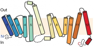
Using additional accessory proteins to help with the orientation of MFSD2A, the authors obtained high-quality images, with a resolution of 0.3 nanometer, or 0.3 billionth of a meter. From the imaging data, the authors found that MFSD2A protein itself is about 5 nm wide and 8 nm long. MFSD2A is a transporter protein and like many transporters, it contains repeated bundles of helices made of protein chains that traverse the cell membrane and are connected by a protein chain that loops within the space in the cell. 

Structure of MFSD2A arranged as protein helices (colored cylinders) within the cell membrane along with protein loops that form both in the extracellular space (“Out”) and within the interior, cytoplasmic space (“In”) of the cell. The cytoplasmic loops likely have an important functional role. Figure from Cater et al, 2021.

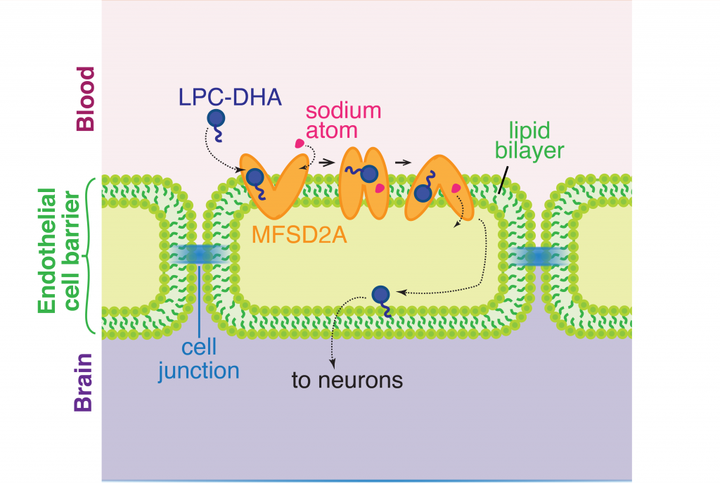
The cell membrane consists of two layers of lipid molecules, known as the lipid bilayer, that allow entry and exit of materials from the cell. These loops provide the shape to the protein inside the cell such that it appears to provide a large enough cavity opening from the lipid bilayer into the cellular space to allow the target molecules to enter the cell. Amino acids are the building blocks of proteins and the cavity contains amino acids of both water-attracting and water-repelling kind. This property makes it possible for many molecules of differing chemical nature to be able to be accommodated within the cavity. This cavity contains three important regions that allow for the protein to be specific and functional: a charged region, a binding site for sodium atoms and a lipid-specific pocket. The authors speculate that these parts help in establishing the mechanism by which LPC-DHA is transported from the outside into the cell. The multiple protein helices form two protein domains that capture LPC-DHA from outside the cell layer of the blood brain barrier of endothelial cells, then rock over a rotation axis so that now their confirmation switches and finally, they release the protein molecule into the cell. For this activity of movement of LPC-DHA, sodium atoms are absolutely required to allow for the shape change of the protein. Once LPC-DHA enters the barrier cells in this manner, the protein is then transported across to the other side of the cell facing the brain containing neurons. 

The transporter channel MFSD2A changes its shape once it binds sodium atoms in the extracellular space, which helps the transport of LPC-DHA from the blood into the brain space through the barrier of a single line of cells made up of endothelial cells. Figure adapted from Cater et al, 2021.

Humans with mutations in MFSD2A gene have abnormal brain defects such as microcephaly, and disruption of the gene in mice affected neuronal branching and fatty acid composition in the brain. The discovery of the structure of a molecule that mediates uptake of essential nutrients across the blood-brain and eye-brain barriers will help in the delivery of therapies of neurological diseases.

Dr. Rosemary J. Cater is a postdoctoral researcher in the lab of Dr. Filippo Mancia in the Department of Physiology and Cellular Biophysics at Columbia University.

Follow this blog

Get every new post delivered right to your inbox.