Contrary to what many people think, bone is a highly dynamic tissue that is constantly being broken down and reformed in order to maintain a healthy and strong skeleton. This process of bone remodeling is enabled by specialized bone cells called osteoclasts and osteoblasts. Osteoclasts produce enzymes to degrade old and damaged bone, which is replaced with new bone by osteoblasts. However, these cells do more than simply breaking down and rebuilding your bones. Recent advances in bone biology have shown that bone cells also have an important endocrine function, meaning that they release hormones into the circulation to affect other tissues and organs in the body. As such, the bone-derived hormone osteocalcin was shown to promote muscle function in a mouse model. Dr. Subrata Chowdhury from the Karsenty lab of the Department of Genetics and Development at CUMC followed up on this remarkable finding, and investigated the regulation of osteocalcin in animal models as well as humans, as recently published in the Journal of Clinical Investigation.
Dr. Chowdhury and colleagues found that circulating osteocalcin levels are increased after a 12-week exercise program in humans, and that this effect requires the signaling molecule, or “cytokine”, interleukin-6 (IL-6). The latter was shown by inhibiting IL-6, which completely blocked the induction of osteocalcin by exercise. They continued by using a mouse model to show that IL-6 is actually derived from the muscle itself, and that its production is necessary for maximal exercise capacity. In other words, mice that could not produce IL-6 in their muscles were not able to run as far on a treadmill as compared to mice that were able to produce IL-6.
They further investigated the interplay between IL-6 and osteocalcin in mice, and found that IL-6 stimulates osteoblasts in the bone tissue to produce RANKL, a protein that is necessary for osteoclast differentiation. As a result, more active osteoclasts are formed within the tissue. These osteoclasts produce high amounts of osteocalcin, which signal back to the muscle to promote the uptake and breakdown of glucose and fatty acids by muscle cells. In addition, osteocalcin stimulates the muscle to produce more IL-6, thereby generating a positive feedback loop between muscle and bone (see Figure below). The end result of this loop is a muscle tissue which can utilize more nutrients from the circulation, and is therefore more functional during exercise.
Exercise capacity, also referred to as fitness, is a strong predictor of chronic disease and mortality. The research by Dr. Chowdhury and colleagues has shown that exercise capacity can be improved by stimulating the IL-6-osteocalcin axis. Although their findings are very convincing, according to Dr. Chowdhury the scientific community initially reacted with disbelief. IL-6 is classically known as an inflammatory cytokine, and is one of the components of the detrimental “cytokine storm” that occurs during, for example, a COVID-19 infection. However, while the high levels of IL-6 under pro-inflammatory conditions are damaging for the body, low sustained levels of IL-6 may actually be beneficial. Follow-up studies are now being performed with low doses of long-acting IL-6 analogues, to study their potential to safely and effectively promote exercise capacity and improve health.
Dr. Chowdhury showed us the importance of not being led by scientific biases, but by our observations. And who would guess that our skeleton does not weigh us down, but actually makes us run faster?
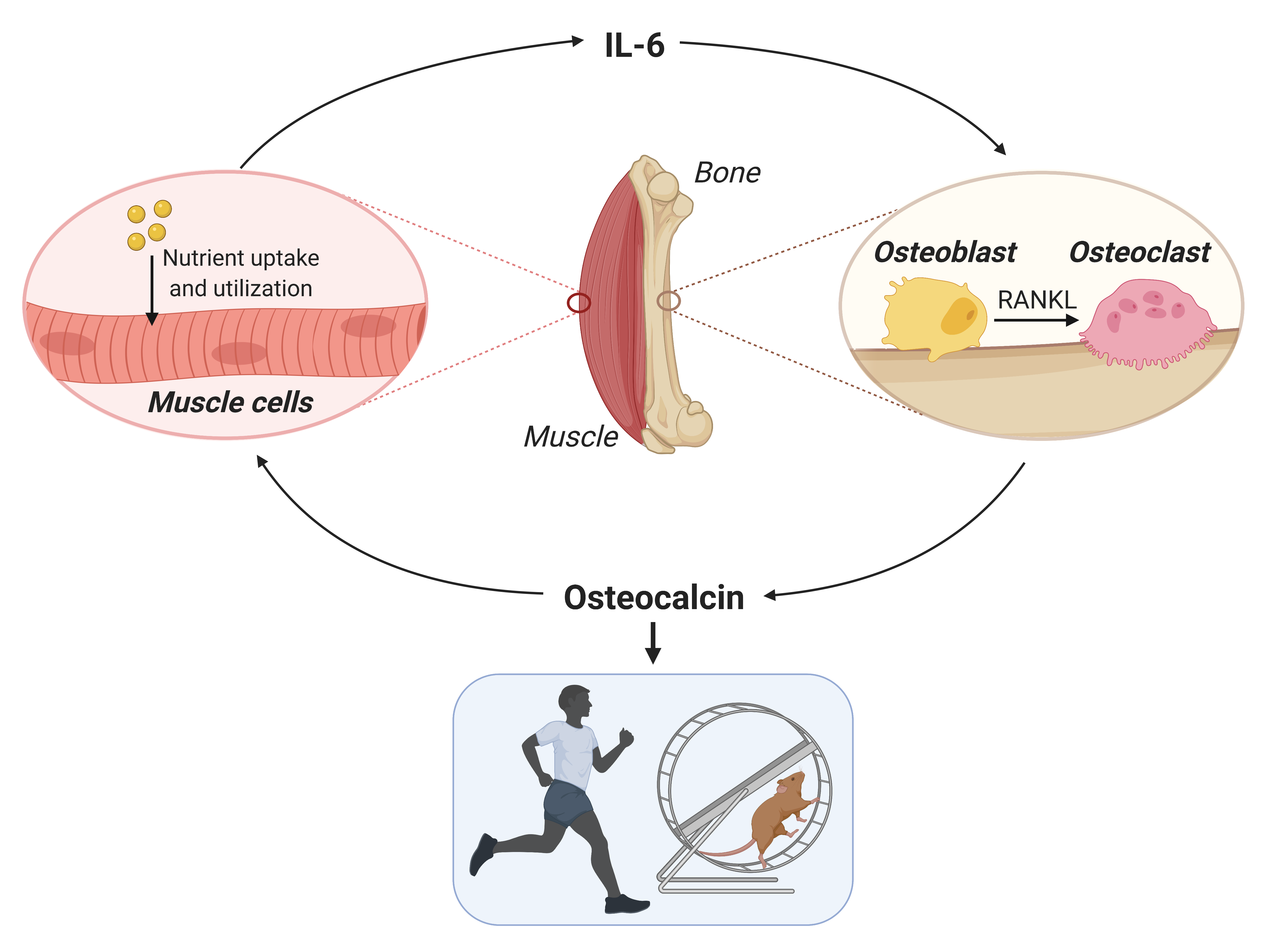 Figure adapted from Chowdhury, JCI 2020, and created with BioRender.com.