Newborn octopus neurons steadily march towards maturity from around the eyes into the brain

Anyone who watched the movie Arrival would not miss the conspicuous resemblance of the alien Heptapods to some of our own earthly beings – the octopuses. While serving as inspiration for alien creatures in movies, as clairvoyants in soccer World Cup or as savages in classic science fiction and mythology, cephalopods like octopuses and squids have been dubbed as one of the most intelligent creatures on the planet. There is a good reason for why cephalopods, particularly octopuses, have developed such a reputation. Octopuses have a striking organization of brain structure, different from that of any other studied organism. They have the largest nervous system among animals lacking a backbone, comprising a total of nine “brains”. Out of these, one is a major donut-shaped brain that contains ~200 million neurons surrounding the octopus’s food pipe, which is strangely located in the head! This brain communicates behavioral intricacies to the eight so-called mini-brains located within the arms, each containing ~40 million neurons. The central brain is responsible for executing complex behaviors like tool use, ability to plan for the future, shape-shift, camouflage, recognize individuals and solve complex puzzles. While the last common ancestor between octopuses and humans was about 680 million years ago, a recent surprising discovery showed that they both evolved to use the same molecules during development. Scientists discovered that genes that produced the camera-like eye in humans are the same ones that gave rise to the camera-like eye in octopuses. What’s more, these cephalopods have evolved complex brains that show behavioral innovation on par with a small primate.
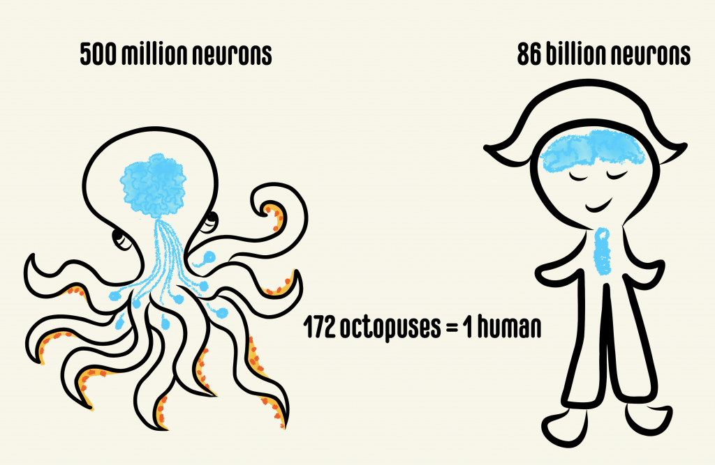
Comparison between the number of neurons present in the octopus and human brains. The octopus has one major brain and eight “mini-brains” while humans have neurons in the head and the spinal cord.

Despite the potential that the octopus provides for understanding developmental biology, particularly the brain, the molecules that dictate how the mollusk’s brain is built are unknown. The common octopus, Octopus vulgaris, is specifically suited to address this question because it can produce thousands of small and transparent eggs in a single batch and scientists have recently mapped out most of its genes. Using O. vulgaris in studies led by Dr. Astrid Deryckere from Dr. Eve Seuntjens’s lab at KU Leuven in Belgium, the group set out to unravel these molecular mysteries. “If you would think of cephalopods as the primates of the sea, that have evolved a complex nervous system from a far more simple ancestral nervous system, surprisingly little is understood on the morphological and molecular mechanisms driving its development.”, said Dr. Deryckere. They approached the problem in two studies. In the first study, she established a system for controlled embryonic development, which enabled her to care for thousands of eggs without the need for the mother octopus. She used state of the art microscopy and recorded high-resolution images of octopus development from fertilization through hatching. This work from Dr. Deryckere and colleagues can now be used as an elaborate reference for cephalopod embryology. 
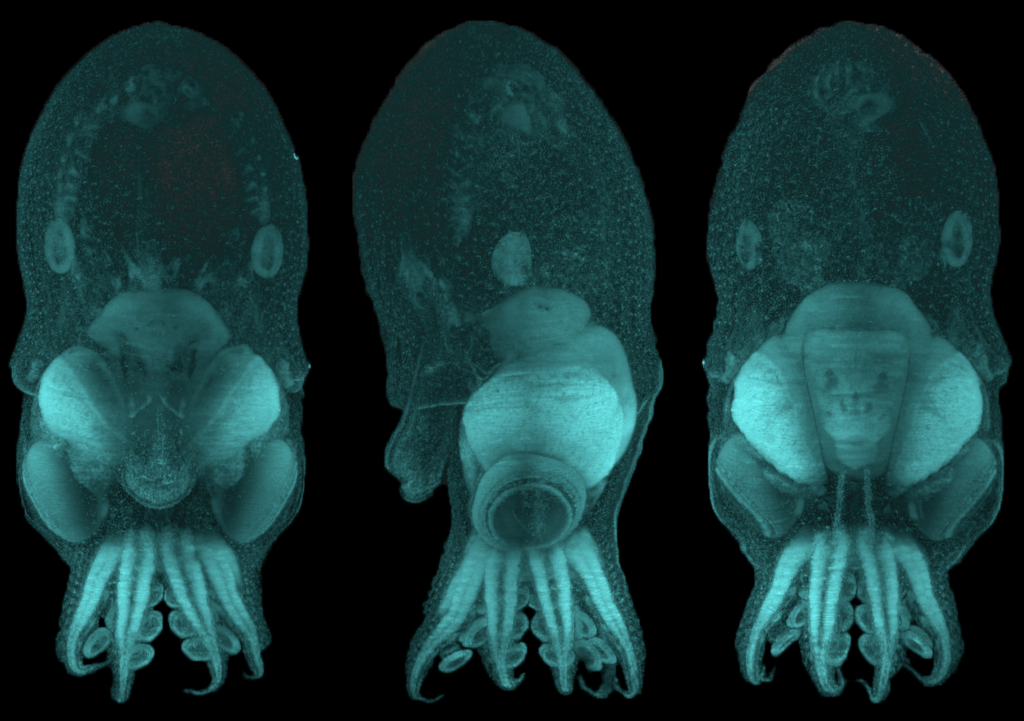
O. vulgaris hatchling imaged in 3D at high resolution after labelling DNA (cyan). The same embryo was imaged from different orientations: back view (left), side view (middle) and front view (right). The head is located on top and the arms are at the bottom. Images were produced by Dr. Astrid Deryckere in the lab of Dr. Eve Seuntjens, KU Leuven, Belgium.

In the second study, Dr. Deryckere dug deep into the origins of the octopus brain. She used precise staging to track the precursor cells of neurons, or “neuronal progenitors”, that generate specialized neurons. Intriguingly, these progenitor cells appeared in structures called the lateral lips, that are unrelated to the brain and are located around the eyes. So, it appeared that neurons were first born in these structures and eventually migrated into the central brain – a possibility that prompted the authors to investigate further. They found that hundreds of thousands of neurons were created within octopus embryos even before hatching. To find out what genes are required for this unique way of making neurons, Dr. Deryckere used molecular markers and showed for the first time that newborn neurons travel long-distances to reach their final location in the brain. The results showed that the genes were expressed in the same order that vertebrates like humans do. By closely observing entire embryos in three dimensions during their growth, she found that neurons proceed through maturation while migrating from the lateral lips to an intermediate transition zone that finally leads to the brain. 
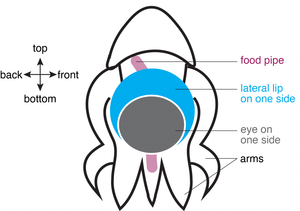
Schematic of O. vulgaris embryo, indicating the location of the lateral lip in relation to the food pipe and the eye. Schematic adapted from images in Deryckere et al., 2021.

Using detailed molecular studies, the scientists now show support for the lateral lips harboring newly dividing neuronal cells in the embryo. This is unusual because unlike in human brains and many other organisms, the dividing cells are located outside the central brain. These dividing cells then unwaveringly make their way towards the final destination to maturity in the octopus central brain. “The migration is especially exceptional for invertebrates where neurons usually migrate only a few cell lengths”, noted Dr. Deryckere about the rarity of the observation.

This unique development in the octopus head and its interesting age-dependent arrangement of dividing cells and mature neurons only inspires further reverence for the cephalopod. While it continues to influence characters in pop culture, the glorious octopus and its brain hold even more promise in the real world. The octopus brain’s cognitive ability has galvanized a new age of artificial intelligence, leading to the construction of flexible robotics and prosthetics, but at the same time, is pushing scientists and philosophers to tackle the important question of how an intelligent life form is defined.

Dr. Astrid Deryckere is currently a postdoc in the lab of Maria Tosches in the Department of Biological Sciences at Columbia University. Her focus remains on brain development but she has transitioned to working on an animal with a backbone – the salamander.
Maternal Stress and the Developing Brain

As humans, we all experience stress. It is a normal, and sometimes even beneficial, part of life. A small amount of stress can help motivate someone to prepare for a job interview or study for an important exam. There are times, however, when stressors become too overwhelming and even detrimental to health. Scientists, from medical researchers to psychologists, have studied stress for decades and documented some of these negative impacts on the brain. When thinking about the importance of the foundational, early years of a person, the presence or lack of stress can play a crucial role in development. For instance, extensive research shows that living in poverty is extraordinarily stressful for families and can negatively influence children’s brain development. The impacts of stress resulting from situations such as growing up in poverty warrant further investigation, especially considering that in 2020, one in six children in the U.S. was living in poverty.

Researchers can use various methods to assess how factors like stress impact the brain of growing children. Developmental scientists can use a tool called EEG, short for electroencephalography, to study the brain. EEG measures electrical activity in the brain by recording the communication between brain cells. It is an ideal neuroimaging method for understanding infant brain development since it allows for infants to be awake and moving, and even sitting on their caregiver’s lap during recording. Besides being infant-friendly, EEG is a useful tool for looking at brain development, given that there is a known pattern of how brain activity changes across the first few years of life.

Specifically, when using EEG to look at brain development, scientists typically see two different patterns. Broadly, infants have a mix of different types of brain activity that we call low-frequency and high-frequency power. Low-frequency power (e.g., theta) tends to be higher when the brain is at rest, while high-frequency power (e.g., alpha, beta, and gamma) tends to be used for more complex thinking like reasoning or language. As infants grow, scientists see that low-frequency power decreases and high-frequency power increases. Importantly, we can use EEG to assess how factors like stress impact the tradeoff of low-frequency and high-frequency power in the developing brain.

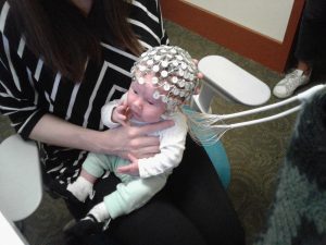
Image of a one-month-old infant with an EEG cap.
Figure 1. A one-month-old infant with an EEG cap. Courtesy of the Neurocognition, Early Experience and Development Lab.

Research shows that children growing up in chronically stressful environments often show alterations in the typical pattern of brain activity development. To further understand the mechanisms underlying this pattern of development, scientists have begun to study which biological and environmental factors may be at play. For instance, researchers can examine the role of caregiver stress, socioeconomic status, home environment, and neighborhood factors, just to name a few.

A recent paper by Dr. Sonya V. Troller-Renfree and colleagues examined maternal stress by looking at the amount of stress hormone (cortisol) found in hair. This measure assesses chronic stress and provides researchers with the average cortisol level of the mother from the preceding 3 months. Dr. Troller-Renfree’s research group hypothesized that infants who have mothers with higher stress hormone, compared to mothers with lower levels of stress, would show differences in their brain activity. Specifically, the researchers predicted that infants of more chronically stressed mothers would exhibit proportionally more low-frequency power and proportionally less high-frequency power compared to infants with physiologically less-stressed mothers.

Indeed, their results showed that infants of mothers who had higher levels of hair cortisol demonstrated higher levels of low-frequency (theta) activity and lower levels of high-frequency (alpha and gamma) brain activity. This finding is consistent with previous research showing that stress and adversity impacts early neural development. Importantly, Dr. Troller-Renfree’s team sampled a diverse pool of participants (both in terms of socioeconomic status and race), therefore bolstering the generalizability of their findings.

So what are the implications of these alterations? Research suggests that similar patterns of neural activity are associated with negative outcomes later in a child’s life, including language development and psychiatric problems. Nevertheless, this does not mean that a child will undoubtedly experience these issues. Additionally, it may be possible that these patterns, while associated with negative outcomes in some areas, may also be adaptive in other circumstances. Furthermore, the issue of the mechanisms by which a mother’s stress impacts the developing child still remains unclear. How exactly does a mother’s stress level impact the brain of her child?

Based on previous research by other scientists, Dr. Troller-Renfree posits a few mechanisms that must be further explored. For example, it is possible that stress impacts crucial mother-child interactions. It could be that stress hormones are passed from mother to baby in utero or through breastmilk. Moreover, it is also possible that environmental factors impact stress and brain development.

It is crucial that developmental scientists continue studying these mechanisms so that targeted intervention programs can be formed for families facing stress. Indeed, the esteemed pediatrician and researcher Dr. Jack Shonkoff of the Center on the Developing Child said in an episode of The Brain Architects Podcast: “In fact, one of the cardinal principles of the science of early childhood development is that if we want to create the best kind of environment for learning and healthy development for young children, we have to make sure that the adults who care for them are having their needs met as well.” As a society, we must recognize how detrimental stress can be to the developing child and invest in finding effective ways to alleviate caregiver stress.

Dr. Sonya V. Troller-Renfree is a Goldberg Postdoctoral Fellow in the Neurocognition, Early Experience and Development Lab at Teachers College, Columbia University. Her research focuses on the effects of early adversity and poverty on cognitive and neural development. She intends to continue examining these questions as part of her new, federally-funded Pathway to Independence Award (K99/00). You can stay up-to-date on her research findings on Twitter at @STRscience or on her website: www.sonyatrollerrenfree.com.

Follow this blog

Get every new post delivered right to your inbox.