Seeing more of the Unseen – using vibrational contrast and MARS to improve microscopy

Biomedical imaging is an important tool in science because it allows scientists to see what may not be visible to the human eye. Using light within the visible spectrum, microscopy allows us to see cells and their functional subunits called organelles, which can be thought of as the internal organs of a cell. We can also visualize certain proteins that may be expressed within certain organelles using fluorescence microscopy. With fluorescence microscopy, proteins in tissue or cells are tagged with light emitting markers, called fluorophores. Fluorophores make proteins under the microscope light up like fireflies on a dark summer night. Different color fluorophores can be used simultaneously to image different proteins at once, however this is limited by the number of colors available in the visible light spectrum. This means that with fluorescence imaging on a confocal microscope, there are a limited number of proteins that can be imaged within a given sample. 

That’s where MARS comes in (not the planet!)! Manhattan Raman Scattering (MARS) is a special dye pallet that, combined with signals from an electronic pre-resonance Stimulated Raman Scattering microscopy (epr-SRS), creates a very sensitive way to probe, visualize and image organelles with vibrational contrast, as opposed to just light contrast. Vibrational contrast detects molecules based on their chemical properties. For example, if a probe molecule has a double bond, it will have a different vibrational frequency than a molecule with a single bond. With this technology, you can differentiate cellular targets by using dyes/probes that vary by light and vibrational signals, making these techniques very sensitive. However, these MARS dyes are difficult to chemically synthesize, and there were initially only a limited number of usable MARS dyes.

Columbia postdoc Dr. Yupeng Miao and colleagues published an article in 2021, summarizing their development of new MARS dyes that have different properties that are easier to synthesize and can visualize even more of the cell’s proteins under the microscope at once! The research contributed 30 new MARS probes that can specifically label various proteins of interest within a given sample.

Before synthesizing these new MARS probes, the researchers designed and simulated models for each potential dye. For the design, they used a similar foundation to the previous MARS probes, but included some adjustments like changing the core atom or substituting stable isotopes throughout the molecule. The results from the design models gave the researchers confidence that they could synthesize these edited molecules to expand the list of available MARS probes.

Indeed, they expanded the list of probes by developing 30 new molecules that are able to label specific cell organelles and functions. For example, MARS probes were used to image subcellular structures including the protein alpha-tubulin, which is a protein within microtubules that provide structural support to the cell, as well as fibrillarin, which is a protein that is used as a nucleoli marker. MARS probes were also shown to successfully target the cell membrane, mitochondria, lysosomes, and other lipid structures within the cell. Even more exciting – this technology allows researchers to probe each of these cellular structures simultaneously, moreso than can be done with current fluorescent microscopy methods. This means that the new MARS probes can be used to image multiple cellular markers within the same sample!

With this technology, scientists can now see even more of the unseen, which can expand our knowledge on cellular (dys)function in health and disease.

Edited by: Maaike Schilperoort, Trang Nguyen

Cleaning Up Data to Spruce Up the Results

Drawing conclusions from scientific studies can be difficult, in part because the data collected may be biased, which leads to a misinterpretation of the data. Let’s say we’re collecting data to investigate how many hours of sleep people get per night, during the week compared to over the weekend. We can ask 100 people their average nightly sleep time on weeknights and on weekends. To avoid bias, or skewing the data toward a particular duration, we should control for a few different factors. For example, we can limit our sample to only ask people 18 years or older, to avoid surveying children who tend to require more sleep than adults. This will avoid introducing a bias in the hours slept per night measure and prevent a trend in the data towards >8 hours a night. 


Some biases cannot be totally avoided during data collection. The existence of this unavoidable bias motivates scientists to consider including confounding variables in their data collection. Scientists use covariates when additional variables that change or differ across groups cannot be controlled for. A covariate is a variable that changes with the variable of interest, but isn’t of particular interest or importance for the question at hand. In our example, there are some other variables that may affect the amount of sleep an adult gets. This can include age (a postdoc in their late 20’s with a grant deadline might not get as much sleep as much as a retiree in their 60’s), activity level (strenuous physical activity leads to more sleep for better recovery), and caffeine intake (maybe serial coffee drinkers sacrifice an extra hour of sleep for an extra large cup in the morning). Because these variables may be different for each participant, we can measure them as observed covariates and include them in our statistical analysis.


Sometimes, as in the case with many epidemiological or public health studies, it’s difficult to measure or control for these covariates because the studies commonly use observational data from population-based studies which might not measure all potential covariates. In these studies, there may be unmeasured biases in the data that produce confounds, leading to imperfect conclusions in population studies. In our example, maybe we neglect to measure time spent on social media, which can affect someone’s total sleep time (I can’t be the only one who scrolls instagram instead of going to sleep at night…). Time spent on social media would be our unobserved covariate, which contributes to unmeasured bias in our sample. 


One way to address the problem of unmeasured bias is to pre-process the data – to fine-tune or clean up the data after it has been collected, but before statistical analysis is performed. In a recent paper, Columbia postdoc Dr. Ilan Cerna-Turoff and colleagues explored the use of a pre-processing method that can be used prior to data analysis in order to reduce the bias introduced by unmeasured covariates in a dataset. 


The pre-processing method investigated in this study is called “Full matching incorporating an instrumental variable (IV)” or “Full-IV Matching”, which aims to reduce biases between groups and thereby improve the accuracy of study findings. An instrumental variable (IV) is a measured variable that is unrelated to the covariates but is related to the variable of interest. For our example, an IV could how comfortable participants find their bed – something that is related to the time spent asleep, but isn’t related to the age or amount of coffee consumed. 


To apply the Full-IV Matching method, the researchers define an IV and “carve out” moderate values of the variable to focus on the extreme values (highest and lowest) across the range of IV measures, essentially ignoring the center of the data set. With this abridged dataset, the researchers implement a “matching” algorithm that pairs individuals who have similar values in their covariates, but who do not have similar values in their IV. In our example, participants who have similar caffeine intake levels or similar ages would be paired with participants who have the opposite bed-comfort level. This explores how the biases in the dataset change when each measured covariate is individually controlled for. Additionally, the researchers can define how much weight should be given to the unobserved covariate, depending on how much bias may be introduced into the data by this unobserved covariate. 


As proof-of-concept, Dr. Cerna-Turoff and colleagues simulated data from a scenario based on the Haitian Violence against Children and Youth Survey. Specifically, data were simulated based on measurements of social characteristics and experiences of young girls in Haiti, who were displaced either to a camp (“exposure” group) or to a wider community (“comparison” group) after the 2010 earthquake. The goal of this simulation experiment was to better understand how the displacement setting may be associated with risk of sexual violence. The researchers simulated data for 5 baseline covariates based on results from the Haitian Violence against Children and Youth Survey: (1) status of restavek (indentureship of poor children for rich families), (2) prior sexual violence, (3) living with parents, (4) age, and (5) social capital, of which the latter is an unobserved covariate. They also generated data for an exposure (camp or community), an outcome (sexual violence against girls), and an IV (earthquake damage severity). The researchers explored how the outcome was affected by the covariates and IV by quantifying the standardized mean difference of the variable across the exposure and comparison groups. A standardized mean difference value close to 0 indicates that the value of the variable was not different across the two groups, suggesting that this variable is not introducing bias into the analysis of group differences. 


The results suggest those who were displaced to a camp were at a higher risk of sexual violence than those who were displaced to a wider community, when correcting for all observed covariates. Additionally, the method successfully balanced the groups when correcting for the unobserved covariate of social capital. If not corrected for, differences in social capital might have confounded these results, such that girls with a stronger support network may appear to be at a lower risk. However, using the Full IV Matching method, bias across exposure and comparison groups for the observed covariates and the unobserved covariate of social capital was reduced, suggesting that neither the social capital nor the observed covariates contributed to the difference in risk for sexual violence observed between the two groups. 


This study provides a proof-of-concept for a pre-processing method for reducing bias across a data set. The authors mention limitations including the effect of the method on sample size and the ‘bias-variance trade-off’, in which increases in accuracy (less bias) may lead to more noise (higher variability) in the data. Ultimately, this type of methodology can aid in the correction of both observed and unobserved biases in population-based data collection, which has significant implications in epidemiologic studies, where not all sources of bias can be measured effectively.


Edited by: Emily Hokett, Pei-Yin Shih, Maaike Schilperoort; Trang Nguyen

Alcohol Use Disorder – are we making the right diagnosis?

Do you and your friends enjoy the occasional cocktail or two over the weekend? Maybe we know someone who enjoys the more-than-occasional cocktail. But, at what point do our drinking habits significantly affect our health? Recent studies suggest that 6% of adults in the United States report heavy or high-risk consumption of alcohol, which is defined as an average of more than 7 drinks/week for women and more than 14 drinks/week for men. This high risk-consumption may lead to Alcohol Use Disorder (AUD) if it is repeated for one year or more. AUD is associated with a number of medical and psychiatric problems, and can even increase risk of death in patients who have cancer and cardiovascular disease.

To diagnose AUD, medical and mental health professionals use the 5th edition of the Diagnostic and Statistical Manual of Mental Disorders (DSM-5), which explores 11 criteria, including alcohol-related cravings, strains on relationships caused by alcohol use, feeling unable to cut back or stop drinking, dangerous or risky behavior when under the influence of alcohol, etc. Unlike previous versions of the DSM, these AUD diagnoses are divided based on severity, where people who experience 0 or 1 of the diagnostic criteria do not have AUD (no-AUD), 2-3 criteria have mild AUD, 4-5 criteria have moderate AUD, and 6+ have severe AUD. However, it’s not well understood whether other factors like the extent of alcohol use, the degree of cravings or impairments, etc. can help classify mild, moderate, and severe AUD diagnoses. 

Last year, Dr. Zachary L. Mannes, a postdoc in the Department of Epidemiology at Columbia University Mailman School of Public Health and New York State Psychiatric Institute, and colleagues published a study in which they aimed to explore any potential relationships between the severity of AUD (no-AUD, mild, moderate, or severe, based on the DSM-5) and self-reported measures of other factors or “external validators”, such as levels of alcohol craving, functional impairment, and psychiatric conditions. To do this, they collected AUD diagnosis as well as measures of external validators in 588 participants. These validators included alcohol specific validators (i.e. Craving, Problematic Use, Harmful Use, Binge Drinking Frequency), psychiatric validators (i.e. Major Depressive Disorder/MDD and posttraumatic stress disorder/PTSD), and functioning validators (social impairments; physical and mental impairments).

Dr. Mannes and colleagues reported that in this cohort of subjects, participants with alcohol use validators had a significantly greater likelihood of a diagnosis with mild, moderate, and severe AUD than a no-AUD diagnosis. Psychiatric validators like MDD and PTSD had a significantly greater likelihood of a severe AUD diagnosis than no-AUD; this relationship was not seen for either mild or moderate AUD. Participants who had social, physical, and mental impairments had a greater likelihood of having severe AUD than no-AUD, but this was not seen for participants with mild or moderate AUD. When looking within participants with an AUD diagnosis (i.e. excluding a no-AUD diagnosis), participants with many alcohol-specific, psychiatric, and functional validators were more likely to have a severe AUD than either mild or moderate AUD.

Overall, the results of this study support the structure of the DSM-5 diagnosis for AUD, as those diagnoses with mild and moderate AUD had significant associations with alcohol use validators, while those with severe AUD had significant associations with alcohol use, psychiatric and functional validators. In other words, people with severe AUD had a higher likelihood of symptoms that affected other aspects of their lives including impairments in social functioning and presence of psychiatric conditions including MDD and BPD. This study emphasizes the importance of looking at levels of severity in AUD as the current DSM-5 does, as opposed to a binary yes/no diagnosis as older versions of the DSM had incorporated. This study also helps further the understanding of optimal ways to diagnose AUD and may help better understand potential treatment implications for various AUD severity. The study published by Dr. Mannes and colleagues supports and progresses the field of AUD research in order to better understand and characterize the symptoms, comorbidities, and diagnosis of AUD, so that medical professionals can better assist those who are struggling with the disorder. 

Edited by: Trang Nguyen, Maaike Schilperoort

Better Work Environments Make Super Nurses Even More Super!

We might all be familiar with the term “burnout” – the feeling of emotional exhaustion or feeling cynical or ineffective with respect to productivity at work, or in relationships with colleagues or clients. The World Health Organization classifies burnout as an occupational, not personal, phenomenon. Studies suggest that burnout can result from poor work environments – not necessarily dependent on the content of the work itself, but instead the setting in which the work is completed and how the work is managed or distributed. Burnout can be prevented or resolved by improving work environments.

Because it is dependent on the environment, the rate of burnout may vary between different job settings. For example, studies suggest that around 40% of the Nursing workforce in the United States is burned out. That’s almost half of all nurses! Nurses, along with Social Workers who also have a burnout rate of about 40%, are among the professions with the highest burnout rates in the country. Nurses have a unique position, as their actions and responsibilities at work directly impact the wellbeing of their patients. Because the lives of their patients may depend on it, it is important that nurses are attentive, motivated, and effective while at their jobs. In other words, nurses should not be burned out in order to properly care for their patients. 

To prevent or resolve burnout in nursing, work environments should aloow appropriate autonomy, or the ability for nurses to use their own discretion and depend on their own expertise to respond to patient care issues. Additionally, positive work environments for nurses include having good working relationships with physicians and hospital administration, and have adequate staffing and resources. If an environment does not include these positive factors, then nurse burnout will likely be prevalent in that clinical setting. Additionally, the combination of a poor work environment and burned out nurses is associated with lower levels of patient care quality and patient outcomes.

A recent study by Columbia postdoc Dr. Amelia Schlak explored how nurse burnout is related to patient care, with the expectation that more nurse burnout would correspond with poorer patient outcomes. Additionally, the researchers investigated how the nurse work environment affects the relationship between nurse burnout and respective patient outcomes. The authors expected to see that nurse burnout will have less of an effect on poorer patient outcomes in better work environments.

In order to investigate these relationships, Dr. Schlak and colleagues measured nurse burnout in over 20,000 nurses across 4 states (CA, PA, FL, and NJ) between 2015–2016 by using the emotional exhaustion subscale of the Maslach Burnout Inventory, which quantifies nurse burnout on a scale from 0 to 54, where higher scores correspond to more burnout. On average, the nurse burnout score in the study was 21/54. They also measured work environment using the Practice Environment Scale of Nursing Work Index survey completed by the same nurses. This measurement accounts for environmental aspects like staffing, access to resources, and nurse-physician relations. The researchers ranked the average hospital environment scores into categories of “poor” (bottom 25%), “mixed” (middle 50%), and “good” (top 25%) environments. They found that the degree of nurse burnout was skewed across the hospital quality category, where most (60%) nurses working in good environments ranked among the lowest burnout levels, while more than 50% of nurses working in poor environments ranked among the most burned out. So, better work environments typically means less burned out and more productive nurses! 

The ultimate priority in healthcare work is, of course, the patient! To see how the environment and nurse burnout affects patients, the researchers also collected patient outcome measurements for each hospital such as (1) patient mortality, (2) failure to rescue, or in-hospital mortality after experiencing an adverse event caused by medical treatment, and (3) length of stay, where only patients with length of stay less than 30 days were considered. The authors found that greater nurse burnout was associated with a higher incidence of patient mortality, an increased rate of failure to rescue and a longer patient stay. Nurses who are not burned out, who are energized and effective, tended to have patients that had better outcomes.

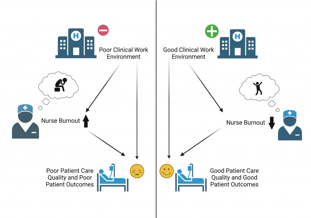
The authors also explored how the nurse work environment affects the relationship between nurse burnout and the patient outcome measurements. When the researchers compared hospitals with poor vs. mixed work environments, as well as mixed vs. good environments, they found that the frequency of burned out nurses decreased, the 30-day in-hospital mortality rate was 14% lower, the failure to rescue rate was 12% lower, and the length of stay was 4% lower in the mixed and good work environments, respectively. This means that by simply improving the work environment (i.e. improving employee relations or providing better resources), hospitals can greatly improve nurse burnout and patient outcomes! This relationship is shown in Figure 1 below. 

Figure 1: Clinical Work environment has an effect on the level of burn out in nurses. Nurse burn out, in turn, has an effect on patient outcomes, where higher levels of burn out result in poorer patient outcomes, and lower levels of outcome result in better patient outcomes. Additionally, the quality of the clinical work environment can also impact patient outcomes, where better outcomes are associated with better hospital environments, while poorer outcomes are associated with poorer hospital environments. Created with BioRender.com

Though this study was based on data from 2015, nurses and other healthcare workers have only become even more burned out in the face of the COVID-19 pandemic, intensified by the overwhelming demand, the pain of losing patients, and the risk of infection that they take every time they go to work. In light of this, hospital management and administration should be proactively addressing healthcare worker burnout, by ensuring that the needs of their healthcare workers are met. This includes, but is not limited to, allowing nurses autonomy or control over their practices, adequate staffing to avoid overworking or long shifts, encouraging and supporting positive relationships among nurses, physicians, and administrative staff, and providing proper resources for nurses to successfully fulfill their responsibilities. 

Also, this past week (May 6th – May 12th, 2022) was Nurses Appreciation Week. Thank you to the Super Nurses for the hard work that you do, oftentimes under relentless and stressful circumstances! You truly are Healthcare Heroes! I hope your hospitals, clinics, or other places of work are prioritizing your work environments, to help reduce the burnout you feel from this pandemic. If they aren’t, send them this article 🙂 

Edited by: Trang Nguyen, Vikas Malik, Maaike Schilperoort

Tau about that! Alzheimer’s protein found in brains of COVID patients

It’s hard not to have COVID on the brain in today’s world – it seems like every conversation ends up on the topic! A recent study completed at Columbia explored the effect of COVID in the brain, by collecting brain samples from the mesial temporal cortex, a brain region implicated in Alzheimer’s disease and responsible for memory, and the cerebellum, a brain region responsible for coordination of movement and balance. Different cellular markers that indicate inflammation and protein build-up in the brain were measured in 10 patients who had passed away from COVID-19 and were compared to brains of those who did not have COVID-19 at the time of death. From this, the researchers were able to infer how COVID-19 infection may alter the brain, potentially causing the neurological symptoms in some COVID patients.

COVID-19 infection can lead to respiratory, cardiac, and neurological symptoms. About one in three COVID patients experience neurological symptoms including loss of taste (hypogeusia), loss of smell (hyposmia), headache, disturbed consciousness, and tingling sensations in their limbs (paresthesia). The exact reason why these neurological symptoms occur is not well understood. In a recent publication, Dr. Steve Reiken and colleagues from the Department of Physiology and Cellular Biophysics at Columbia University Vagelos College of Physicians and Surgeons explore how factors associated with COVID infection, like inflammation, led to these neurological symptoms.

SARS-CoV-2, the virus that causes COVID-19, enters the body through the airways. The spike proteins on the surface of SARS-CoV-2 virus facilitate the entry into cells through the angiotensin converting enzyme 2 (ACE2) receptor. This leads to inflammation in the lungs and other organs. ACE2 receptors are downregulated during COVID infection, a pattern which has been tied to an upregulation of inflammatory marker transforming growth factor-𝛃 (TFG-𝛃) in other disease models including cancer. Lower ACE2 activity has also been tied to greater concentrations of Alzheimer’s disease (AD) related proteins amyloid-𝛃 (A𝛃) and phosphorylated tau. Perhaps the entry-point of the SARS-CoV-2 virus activates inflammation pathways that can affect the brain similar to the way it is affected in AD, and might cause the neurological issues that sometimes come with COVID infection.

In the study, inflammation markers that represent TFG-𝛃 levels were measured in the brain samples of COVID patients and were compared to non-patients. Each of these measures were higher in brain samples of COVID patients, suggesting that COVID infection contributed to more inflammation in the brain.

Inflammation may have downstream effects that can impact the function of healthy tissues. For example, the highly-regulated use of the calcium ion (Ca2+), which is a key player in cell-to-cell communication, can become impaired in conditions of inflammation. Specifically, the ryanodine receptor (RyR) is an ion channel protein which is responsible for Ca2+ release. When in an open configuration, Ca2+ can flow freely through the channel. To stop Ca2+ flow, helper proteins interact with the RyR to stabilize the closed configuration of the channel. Previous studies have suggested that these helper proteins are downregulated in inflammation, which means that the RyR is more likely to be unstable, resulting in excess Ca2+ flow, or a Ca2+ leak. Ca2+ leaks have been thought to contribute to a number of diseases, including the development of tau pathology in AD.

In Dr. Reiken and colleagues’ study, indicators of typically functioning RyR were measured in the brain samples of COVID patients and non-patients. These measures included the amount of RyR channel in the open configuration (which means a lot of free flowing Ca2+) and the concentration of the helper proteins that helps the RyR remain stable in the closed configuration. The researchers found that there were less helper proteins in the COVID brains compared to the non-COVID brains. Additionally, more of the RyR channels were in an open configuration in the COVID brains compared to non-COVID brains. This means that Ca2+ leaks were more likely to happen in the brains of those infected with COVID-19.

In addition to cellular markers of inflammation and Ca2+ leaks, Dr. Reiken and colleagues also investigated levels of AD-related proteins A𝛃 and tau aggregation in the brains of control subjects and COVID patients. For A𝛃, relevant protein levels were similar between COVID patients and controls, suggesting that COVID does not cause the collection of A𝛃 in the brain. However, the concentration of phosphorylated tau, another protein that is highly implicated in AD pathology, was higher in the temporal lobe and cerebellum regions in COVID patients compared to control subjects.

To take this one step further, the researchers treated the COVID patients’ brain samples with Rycal ARM210, a drug that is currently in clinical trials for other applications at the NIH (NCT04141670) and helps to reduce Ca2+ leak. With ARM210, helper protein levels in the COVID brain samples increased from the original levels in the COVID brain samples that were not treated with the drug. Additionally, the amount of RyR in the open configuration decreased in the COVID brain samples with ARM210, compared to the un-treated samples. Thus, treatment with this drug may combat Ca2+ leak in brain tissue. If unstable RyR leads to the Ca2+ leak, and Ca2+ leak can promote tau phosphorylation and build up in the brain, then using the Rycal drug ARM210 to target and limit Ca2+ release may potentially be a way to treat of these brain abnormalities in COVID-19 and possibly minimize neurological symptoms.

Given these results, the authors propose a mechanism by which infection with the SARS-CoV-2 virus may lead to protein aggregation similar to tau deposition in AD. An adaptation of the proposed mechanism is shown in the Figure below.

Figure 1 (above): Proposed mechanism for neurological symptoms of COVID-19 infection. Adapted from Reiken et al., 2022. Created with BioRender.com.

Though this is a very exciting study exploring the neurobiology in COVID brains, there are some additional things to consider. Firstly, while inflammatory markers were elevated in the brains of COVID patients, SARS-CoV-2 virus particles were not detectable in the brain. This suggests that these effects are caused by systemic factors, and are not localized to cells that are infected with SARS-CoV-2. Additionally, in terms of the AD-related proteins, elevated phosphorylated tau protein was detected in the mesial temporal cortex and the cerebellum of COVID patients compared to controls. In AD, tau protein collects in the medial temporal cortex early in disease progression, but does not collect in the cerebellum. This, in addition to the lack of A𝛃 aggregation in the COVID patients’ brain samples, is a marked difference between the pathology of the brain in AD and in COVID. However, distribution and amount of tau protein in AD is linked to cognitive abilities, so perhaps the collection of tau in the brain of COVID contributes to cognitive symptoms like “brain fog”. The current study used brain samples from 10 COVID patients, but did not collect cerebrospinal fluid samples or use animal models to validate these findings yet. Future work that addresses these limitations and further questions may help us fully understand the role of COVID in the brain, and may help with treatments for those who are struggling with prolonged neurological symptoms of COVID.


Dr. Reiken, the first author of this work, is an Assistant Professor at Columbia University Department of Physiology. Dr. Dridi, a Postdoc Fellow at Columbia, and Dr. Liu, a Postdoc Research Scientist at Columbia, also contributed to this work. Find the original research article here.

Reference:
Reiken, S, Sittenfeld, L, Dridi, H, Liu, Y, Liu, X, Marks, AR. Alzheimer’s-like signaling in brains of COVID-19 patients. Alzheimer’s Dement. 2022; 1- 11. https://doi.org/10.1002/alz.12558
Follow this blog

Get every new post delivered right to your inbox.