How a molecular structure explains the transport of fatty acids past the blood-brain barrier

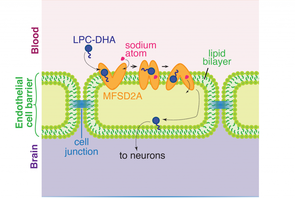
The brain and eyes develop through constant circulation of nutrients through the blood-brain and blood-retina barriers. One such nutrient that is essential for development is an omega-3 fatty acid called docosahexaenoic acid (DHA). DHA makes up a fifth of all the fatty acids required on the membranes of cells in the central nervous system. Neither the neurons in the brain nor the cells in the eye are capable of synthesizing DHA by themselves and therefore depend on dietary sources for DNA. Previously, scientists knew from cellular clues that this fatty acid most likely passed through to the blood-brain and blood-retina barriers in the form of lysophophatidylcholine (LPC-DHA) using a molecular channel. This transporter is known as a major facilitator superfamily domain containing 2A, or MFSD2A, with the help of sodium atoms regulating the channel. However, it was not clear how this channel allowed the passing of complex molecules like DHA. A recent study by Dr. Rosemary Cater and colleagues at Columbia University provided precise clues to further show the structure of this channel. 

To investigate the structure of MFSD2A, the authors used a state of the art imaging technique called single-particle cryo-electron microscopy. This is a method of electron microscopy where a beam of electrons is transmitted through a rapidly frozen purified molecule. Because the sample is flash frozen, the molecules trapped in a frozen state can be imaged in their native shape as present in the cell and from multiple angles. By capturing and combining multiple captured 2D images, a 3D structure of the protein can be reconstructed with extreme accuracy. Cryo-electron microscopy is so impactful in biological significance that this method was awarded the 2017 Nobel Prize in Chemistry. found a number of molecular patterns and arrangements of protein chains that make up a full molecule of MFSD2A

Protein structure studies are typically among the most challenging grounds to explore in biology because proteins need to be captured in their native state as present in the cell. Past discoveries of various protein structures have been so instrumental in shaping therapeutic areas that the extent of mechanistic understanding of biological molecules has resulted in recognition by Nobel committees. Most recently, the discovery of the structure of the ribosome opened up fields of exploration into therapeutic interventions into ribosome diseases, some of which can lead to cancer.

To get the best chance at imaging the structure of MFSD2A, the scientists extracted and examined purified versions of this protein obtained from multiple organisms: the zebrafish, the frog, European cattle, the domestic dog, the red junglefowl, mice and humans. Finally, the authors found that the protein obtained from the red junglefowl, which is a rooster species that originates from Southeast Asia, was the most experimentally stable and most alike (73% identity) the human version of MFSD2A. 

Using additional accessory proteins to help with the orientation of MFSD2A, the authors obtained high-quality images, with a resolution of 0.3 nanometer, or 0.3 billionth of a meter. From the imaging data, the authors found that MFSD2A protein itself is about 5 nm wide and 8 nm long. MFSD2A is a transporter protein and like many transporters, it contains repeated bundles of helices made of protein chains that traverse the cell membrane and are connected by a protein chain that loops within the space in the cell. 

Structure of MFSD2A arranged as protein helices (colored cylinders) within the cell membrane along with protein loops that form both in the extracellular space (“Out”) and within the interior, cytoplasmic space (“In”) of the cell. The cytoplasmic loops likely have an important functional role. Figure from Cater et al, 2021.

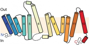
The cell membrane consists of two layers of lipid molecules, known as the lipid bilayer, that allow entry and exit of materials from the cell. These loops provide the shape to the protein inside the cell such that it appears to provide a large enough cavity opening from the lipid bilayer into the cellular space to allow the target molecules to enter the cell. Amino acids are the building blocks of proteins and the cavity contains amino acids of both water-attracting and water-repelling kind. This property makes it possible for many molecules of differing chemical nature to be able to be accommodated within the cavity. This cavity contains three important regions that allow for the protein to be specific and functional: a charged region, a binding site for sodium atoms and a lipid-specific pocket. The authors speculate that these parts help in establishing the mechanism by which LPC-DHA is transported from the outside into the cell. The multiple protein helices form two protein domains that capture LPC-DHA from outside the cell layer of the blood brain barrier of endothelial cells, then rock over a rotation axis so that now their confirmation switches and finally, they release the protein molecule into the cell. For this activity of movement of LPC-DHA, sodium atoms are absolutely required to allow for the shape change of the protein. Once LPC-DHA enters the barrier cells in this manner, the protein is then transported across to the other side of the cell facing the brain containing neurons. 

The transporter channel MFSD2A changes its shape once it binds sodium atoms in the extracellular space, which helps the transport of LPC-DHA from the blood into the brain space through the barrier of a single line of cells made up of endothelial cells. Figure adapted from Cater et al, 2021.

Humans with mutations in MFSD2A gene have abnormal brain defects such as microcephaly, and disruption of the gene in mice affected neuronal branching and fatty acid composition in the brain. The discovery of the structure of a molecule that mediates uptake of essential nutrients across the blood-brain and eye-brain barriers will help in the delivery of therapies of neurological diseases.

Dr. Rosemary J. Cater is a postdoctoral researcher in the lab of Dr. Filippo Mancia in the Department of Physiology and Cellular Biophysics at Columbia University.

The key to a longer life might be in skipping that midnight snack

Have you ever caved in to the temptation of a snack in the middle of the night that manifested into a quick freezer dive to grab that ice-cream or into a series of quick taps on your food delivery app to get those udon noodles? Suffice it to say that I have been a victim to this thought one too many times. Much to my chagrin, there is an abundance of evidence that suggests that eating during restricted hours of the day or time-restricted feeding (TRF) can slow down decline of bodily functions. Limiting food intake to certain hours of daytime, even if the food is not necessarily nutritious or low in calories, can prevent ageing or even kickstart anti-aging mechanisms in mice and flies with obesity or heart disease. Because ageing was dependent on when the body takes in food, these studies hint at the role of the body’s biological clock, known as circadian rhythms, in regulating health and longevity. In an unexpected new study authored by Columbia postdoc Dr. Matt Ulgherait, flies following time-restricted feeding while also balancing it with an unlimited all-access ad libitum diet, show a significant increase in lifespan. 

By structuring 24 hour day-night periods as cycles of 12 hours of light followed by 12 hours of darkness in a temperature-controlled box, the authors tested various dietary regimens for their effects on lifespan and stumbled upon one regimen that consistently showed longer lifespan along with enhanced health in the flies. This regimen cycled between a 20-hour fast starting at mid-morning (6 hours after lights on) to a 22 hour recovery period of eating ad-libitum on repeat in young flies within 10-40 day post hatching stage of adulthood. However, flies that began this regimen after reaching older age at day 40 did not show enhanced lifespan. In comparison to flies that were allowed access to food ad-libitum on a 24 hour cycle, flies following this particular fasting-feeding regimen showed a 18% increase in female lifespan and 13% increase male lifespan in their young age. Due to the cycling schedule of unlimited food access with periods of fasting, the authors termed this regimen as intermediate time-restricted feeding (iTRF).

Previous studies have shown that caloric restriction through reduced food intake, protein restriction or inhibiting insulin-like signaling can extend lifespan. However, iTRF did not appear to limit flies from eating less and in many cases, resulted in flies eating more during times of food access compared to those in the ad libitum group. Thus, lifespan extension under iTRF did not occur because of limitation in nutrient uptake. Interestingly, an iTRF regimen performed under additional treatments of either dietary protein restriction or inhibited insulin-like signaling, resulted in a marked boost in lifespan compared to iTRF alone. It therefore seems that  independent mechanisms that  can enhance lifespan can be combined to increase lifespan even more. 

While these methods provide ways to extend lifespan through incremental means, some might argue that it would be meaningless to simply survive without long-lasting health benefits. To examine whether the longer-lived flies continued to exhibit youth, scientists measured the fitness of the flies using two well-known age-related tests: the flies’ ability to climb up the plastic vial they are in and how much they accumulate in their tissues aggregates of aging proteins – polyubiquitin and p62. When compared to the ad libitum group, iTRF flies climbed much faster and had fewer polyubiquitin and p62 aggregates in the flight muscles, even after they reached an age beyond 40 days of hatching. While the gut microbiome was shown to dictate proclivity for disease and thus have an effect on lifespan, the gut tissue in iTRF flies remained healthier with more normal cells, even when the gut microbiome was depleted with antibiotics. Therefore, the flies appeared to be in optimal health conditions with fewer aging markers in addition to longer survival, demonstrating yet again that aging slowed down due to better functioning of organs.

The dietary regimen under iTRF only controls the timing of feeding but not the nutritional intake, which provided clues to the authors that perhaps the body’s natural biological clock had something to do with iTRF-mediated lifespan. The biological clock in flies consists of proteins that are also present in other organisms all the way from fungi to humans. The main molecular parts of the core circadian clock include the proteins ‘Clock’ (Clk) and ‘Cycle’ (Cyc) which activate the genes period (per) and timeless (tim), which in turn inhibit Clk and Cyc. This process is called a feedback loop which takes all of 24 hours to complete in both flies and humans, and this is how our bodies respond to light-dark cycles. Flies undergoing iTRF showed enhanced expression of Clk in the daytime and of per and tim at night time. The authors then explored the feeding behavior and metabolism of circadian clock genetic mutants undergoing iTRF and found that neither the 20 hour long fasting period nor dietary restriction in their food altered their feeding behavior when compared to normal flies under iTRF. Yet, the extended lifespan was completely missing in Clk, per and tim mutants undergoing iTRF. Even the improved health seen with an iTRF regimen through better climbing ability and less aging-protein aggregation was abolished in per mutants compared to normal flies. Shifting the iTRF cycle by 12 hours with a fasting period during the daytime abolished the occurrence of an extended lifespan. In the altered regimen, while the same cycle was now only shifted by half a day, eating at night time while fasting during the day just did not work. This discovery showed that there could be a deep link between the body’s biological timer and when during the day food is eaten that determines both longevity and well-being. 

Because shifting the fasting period to daytime did not show any benefits, the authors checked whether genes that activate during fasting are also linked to the biological clock. In fact, Dr. Ulgherait and group had already shown that disrupting tim and per genes in the gut, which is where food is processed, caused an increase in lifespan. But, iTRF included periods of starvation that could trigger different metabolic processes. Starvation induces cellular mechanisms to degrade and recycle its molecules in a process called autophagy. Interestingly, genes encoding two autophagy proteins, Atg1 and Atg8a, which are also present in humans, showed peak levels in the night time with enhanced peaks in flies under iTRF. During autophagy, there is an increased activity of cell organelles called lysosomes that contain digestive enzymes needed to break down cellular parts. The authors found that normal flies fasting under iTRF showed higher Atg1 and Atg8a expression along with more lysosomal activity but period mutants failed to do so. Using some more genetic tricks, the authors found that manipulating the level of autophagy to go up or down directly showed an effect on iTRF-mediated lifespan.

Finally, to explore the link between iTRF-mediated lifespan and autophagy, the authors used genetic tools to increase night-specific levels of Atg1 and Atg8a. In a surprising revelation, flies with night-specific expression of Atg1 and Atg8a showed an increase in lifespan, even when these flies did not undergo fasting and were fed ad libitum. Subjecting these genetically altered flies to iTRF did not additionally increase their lifespan, suggesting to the authors that circadian enhancement of cellular degradation under an all-access diet provides the same beneficial effects as fasting done under the stricter regimen of iTRF. Flies with night-specific enhanced autophagy also showed better neuromuscular and gut health on an all-access diet. Therefore, clock-dependent enhancement of the biological recycling machinery can mimic the lifespan extension mediated by iTRF.


Now of course large genetic manipulations are not yet a consideration in humans but this study provides a potentially powerful yet simple change in dietary strategy that could just somehow slow down aging. Aging increases risk of mortality and disease but imagine a food intake regimen translatable from this study into humans that can help improve overall neuromuscular and gut health. So, while technology has indeed made it so much easier than before to have food at our doorstep in a few phone taps in the middle of the night, perhaps restricting the hours of when we eat can really help us live healthier lives. This study now makes me reconsider the famous quote by Woody Allen in the context of food, “You can live to be a hundred if you give up all the things that make you want to live to be a hundred”.

Dr. Matt Ulgherait is a postdoctoral researcher in the lab of Dr. Mimi Shirasu-Hiza in the department of Genetics & Development at Columbia University. Dr. Ulgherait and his colleagues also recently showed that removing the expression of the period gene from the gut tissue was sufficient to cause an increase in lifespan.

Ancestry connects non-cancer causing mutations in cancer patients

The cause of cancer as a disease has been partly attributed to genetics across a diverse range of populations. However, it is unclear whether cancer patients carry additional genetic mutations, also known as variants, in non-cancer causing genes and if these variants are evolutionarily related. Because ancestry-specific variants were more recently generated in evolutionary time, they could have been easily missed in analyses where all patients were cumulatively analyzed without consideration for ancestry. A recent concept proposed by geneticists suggests that people are more likely to develop or be protected from diseases based on recently acquired mutations and are less so due to more distant mutations. This is an interesting theory that scientists can now test using genome information from more than 10,000 cancer patients whose ancestries are known. So far, how mutations affect gene expression – whether they completely abolish the expression of gene products (e.g. protein) or result in the creation of a misshapen protein, have only been reported for variants present in patients with European ancestry. The remaining ancestries are yet to be explored.


Advances in sequencing technology have made it easier for researchers to access genome sequencing information under clinical settings and for healthcare providers to share personalized diagnoses as part of ‘genomic medicine’ to patients. Using publicly available genome sequencing data for cancer patients, Dr. Xiao Fan and colleagues analyzed the variants in non-cancer causing genes and in “medically actionable” genes in 10,389 cancer patients. The authors found 1.46 billion mutations, which were then filtered through rigorous quality testing of sequencing information followed by expert geneticist review, resulting in a final total reliable set of 2,920 non-cancer related pathogenic and likely pathogenic variants. About 750 of these variants were harbored on average within a quarter of the cancer cases, no matter the heritage. A surprising majority (~27%) of the total variants were displayed in patients with European ancestry, followed sequentially by Latinx/Native American (15%), African American (13%) and East Asian (12%) patients.


Because genetic mutations can affect expression of proteins, the authors then dug deep into the variant data to examine whether these variants behaved in an expected manner on a molecular level. When genes contain mutations that cause the protein it encodes to be a shorter version of itself, the mutation is referred to cause a protein “truncation”. Sometimes, a truncating mutation in a gene can trigger a decrease in expression at the messenger RNA (mRNA) level even before the mRNA is used to make the protein. To find out if the variants that produced truncated gene products underwent changes at the mRNA level, the authors measured the gene expression levels of such variants. Of the variants that showed a meaningful difference in gene expression compared to non-cancer patients, a large majority of variants showed a decrease in expression. This result indicated to the authors that truncation-causing variants often work at the mRNA level even before the cells spend energy to make the disease-associated proteins. The authors then examined the behavior of gene variants that do not cause truncations but rather cause just a single swap in the gene sequence, known as “missense” variants. Missense mutations typically only cause a change in one or two building blocks of the protein but do not affect the abundance of the protein itself. Surprisingly, the authors found that the missense variants in their data are unusually regulated in the cancer patients at the mRNA level resulting in a decrease in gene, and therefore, protein expression. This is an uncommon observation, making the authors speculate that missense variants are perhaps controlled by gene-expression independent mechanisms within the cancer patients’ cells.

This study provides a testament to the power of genomic medicine that can be used to complement conventional medical treatment. With a strong sample of ~10,000 cancer patients, this report stands as one of the most comprehensive studies that considers race and ancestry in its analysis. While genomic profiling is becoming more common in medical diagnoses, this study further provides a reason for understanding diseases and invention of medicine based on race, ethnicity and genetic heritage.

Newborn octopus neurons steadily march towards maturity from around the eyes into the brain

Anyone who watched the movie Arrival would not miss the conspicuous resemblance of the alien Heptapods to some of our own earthly beings – the octopuses. While serving as inspiration for alien creatures in movies, as clairvoyants in soccer World Cup or as savages in classic science fiction and mythology, cephalopods like octopuses and squids have been dubbed as one of the most intelligent creatures on the planet. There is a good reason for why cephalopods, particularly octopuses, have developed such a reputation. Octopuses have a striking organization of brain structure, different from that of any other studied organism. They have the largest nervous system among animals lacking a backbone, comprising a total of nine “brains”. Out of these, one is a major donut-shaped brain that contains ~200 million neurons surrounding the octopus’s food pipe, which is strangely located in the head! This brain communicates behavioral intricacies to the eight so-called mini-brains located within the arms, each containing ~40 million neurons. The central brain is responsible for executing complex behaviors like tool use, ability to plan for the future, shape-shift, camouflage, recognize individuals and solve complex puzzles. While the last common ancestor between octopuses and humans was about 680 million years ago, a recent surprising discovery showed that they both evolved to use the same molecules during development. Scientists discovered that genes that produced the camera-like eye in humans are the same ones that gave rise to the camera-like eye in octopuses. What’s more, these cephalopods have evolved complex brains that show behavioral innovation on par with a small primate.

Comparison between the number of neurons present in the octopus and human brains. The octopus has one major brain and eight “mini-brains” while humans have neurons in the head and the spinal cord.

Despite the potential that the octopus provides for understanding developmental biology, particularly the brain, the molecules that dictate how the mollusk’s brain is built are unknown. The common octopus, Octopus vulgaris, is specifically suited to address this question because it can produce thousands of small and transparent eggs in a single batch and scientists have recently mapped out most of its genes. Using O. vulgaris in studies led by Dr. Astrid Deryckere from Dr. Eve Seuntjens’s lab at KU Leuven in Belgium, the group set out to unravel these molecular mysteries. “If you would think of cephalopods as the primates of the sea, that have evolved a complex nervous system from a far more simple ancestral nervous system, surprisingly little is understood on the morphological and molecular mechanisms driving its development.”, said Dr. Deryckere. They approached the problem in two studies. In the first study, she established a system for controlled embryonic development, which enabled her to care for thousands of eggs without the need for the mother octopus. She used state of the art microscopy and recorded high-resolution images of octopus development from fertilization through hatching. This work from Dr. Deryckere and colleagues can now be used as an elaborate reference for cephalopod embryology. 

O. vulgaris hatchling imaged in 3D at high resolution after labelling DNA (cyan). The same embryo was imaged from different orientations: back view (left), side view (middle) and front view (right). The head is located on top and the arms are at the bottom. Images were produced by Dr. Astrid Deryckere in the lab of Dr. Eve Seuntjens, KU Leuven, Belgium.

In the second study, Dr. Deryckere dug deep into the origins of the octopus brain. She used precise staging to track the precursor cells of neurons, or “neuronal progenitors”, that generate specialized neurons. Intriguingly, these progenitor cells appeared in structures called the lateral lips, that are unrelated to the brain and are located around the eyes. So, it appeared that neurons were first born in these structures and eventually migrated into the central brain – a possibility that prompted the authors to investigate further. They found that hundreds of thousands of neurons were created within octopus embryos even before hatching. To find out what genes are required for this unique way of making neurons, Dr. Deryckere used molecular markers and showed for the first time that newborn neurons travel long-distances to reach their final location in the brain. The results showed that the genes were expressed in the same order that vertebrates like humans do. By closely observing entire embryos in three dimensions during their growth, she found that neurons proceed through maturation while migrating from the lateral lips to an intermediate transition zone that finally leads to the brain. 

Schematic of O. vulgaris embryo, indicating the location of the lateral lip in relation to the food pipe and the eye. Schematic adapted from images in Deryckere et al., 2021.

Using detailed molecular studies, the scientists now show support for the lateral lips harboring newly dividing neuronal cells in the embryo. This is unusual because unlike in human brains and many other organisms, the dividing cells are located outside the central brain. These dividing cells then unwaveringly make their way towards the final destination to maturity in the octopus central brain. “The migration is especially exceptional for invertebrates where neurons usually migrate only a few cell lengths”, noted Dr. Deryckere about the rarity of the observation.

This unique development in the octopus head and its interesting age-dependent arrangement of dividing cells and mature neurons only inspires further reverence for the cephalopod. While it continues to influence characters in pop culture, the glorious octopus and its brain hold even more promise in the real world. The octopus brain’s cognitive ability has galvanized a new age of artificial intelligence, leading to the construction of flexible robotics and prosthetics, but at the same time, is pushing scientists and philosophers to tackle the important question of how an intelligent life form is defined.

Dr. Astrid Deryckere is currently a postdoc in the lab of Maria Tosches in the Department of Biological Sciences at Columbia University. Her focus remains on brain development but she has transitioned to working on an animal with a backbone – the salamander.

Follow this blog

Get every new post delivered right to your inbox.